Homeostasis: key idea
· Homeostasis = maintenance of a constant internal environment within narrow limits.
· Important because enzymes and cells work best at optimum conditions.
· In mammals, key variables include core body temperature, blood glucose concentration and blood water potential.
· Stable tissue fluid keeps cells supplied with nutrients and oxygen and removes wastes efficiently.
· Homeostasis prevents harmful changes that could reduce enzyme activity, disrupt cell signalling or damage cells.
Principles of homeostatic control
· A stimulus is a detectable change in the internal or external environment.
· Receptors detect the stimulus and send information to a coordination system.
· Coordination systems = nervous system and endocrine system.
· Effectors = muscles or glands that bring about a response.
· Negative feedback returns the variable back towards its set point / normal range.
· Negative feedback prevents overcorrection by switching off or reducing the response once normal conditions are restored.
Urea production
· Urea is produced in the liver.
· It is formed from deamination of excess amino acids.
· Deamination removes the amino group from amino acids.
· Urea is transported in the blood to the kidneys for excretion in urine.
· Exam link: amino acids cannot be stored, so excess amino acids must be deaminated.
Kidney structure
· Human kidney structures to know: fibrous capsule, cortex, medulla, renal pelvis, ureter, branches of renal artery and branches of renal vein.
· Fibrous capsule = tough outer covering that protects the kidney.
· Cortex = outer region containing glomeruli, Bowman’s capsules, proximal convoluted tubules and distal convoluted tubules.
· Medulla = inner region containing loops of Henle and collecting ducts.
· Renal pelvis = funnel-shaped space collecting urine before it passes into the ureter.
· Renal artery supplies blood to be filtered; renal vein carries blood away after filtration and reabsorption.
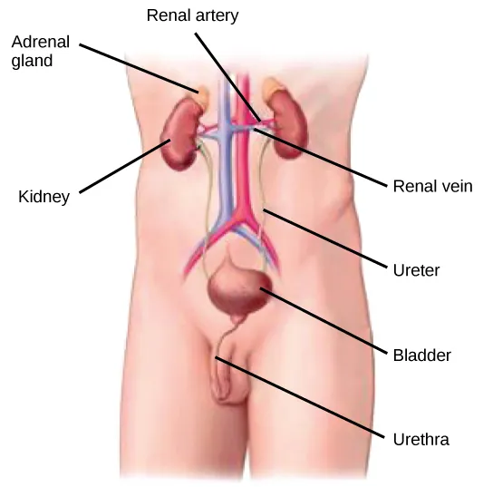
This diagram shows the main regions of the kidney and helps connect kidney anatomy to urine formation. The cortex and medulla contain different parts of the nephron. The renal pelvis collects urine before it enters the ureter. Source
Nephron parts to identify
· A nephron is the functional unit of the kidney.
· Parts to identify in diagrams, photomicrographs and electron micrographs: glomerulus, Bowman’s capsule, proximal convoluted tubule, loop of Henle, distal convoluted tubule and collecting duct.
· Associated blood vessels to recognise: afferent arteriole, efferent arteriole, glomerular capillaries and capillaries surrounding the tubule.
· Glomerulus = capillary knot where blood is filtered.
· Bowman’s capsule = cup-shaped structure that collects glomerular filtrate.
· Proximal convoluted tubule = main site of selective reabsorption.
· Collecting duct = site where water reabsorption is adjusted during osmoregulation.
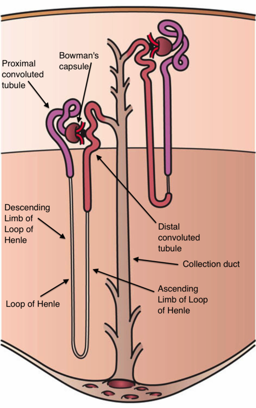
This diagram shows the major parts of a nephron in sequence. It is useful for learning where ultrafiltration, selective reabsorption and water regulation occur. Focus on linking each labelled region to its function. Source
Formation of urine: ultrafiltration
· Ultrafiltration occurs in the Bowman’s capsule.
· Blood enters the glomerulus through the afferent arteriole and leaves through the efferent arteriole.
· The afferent arteriole is wider than the efferent arteriole, producing high hydrostatic pressure in the glomerulus.
· High pressure forces small molecules out of the blood into Bowman’s capsule to form glomerular filtrate.
· Filtrate contains water, glucose, amino acids, urea and mineral ions.
· Large molecules and cells remain in blood, including red blood cells, white blood cells, platelets and plasma proteins.
Bowman’s capsule: structure and function
· The filtration barrier includes capillary endothelium, basement membrane and podocytes.
· Capillary endothelium has pores that allow plasma and small solutes through.
· Basement membrane acts as a molecular filter, preventing large proteins from passing through.
· Podocytes have foot-like projections with gaps called filtration slits.
· These adaptations allow rapid filtration of small molecules while retaining cells and large proteins in the blood.
· Exam phrase: high hydrostatic pressure forces small molecules through the filtration barrier into Bowman’s capsule.
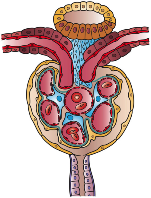
This image helps explain the cellular structure involved in ultrafiltration. The renal corpuscle is specialised to allow small molecules to pass into the filtrate while larger blood components remain in the capillaries. It is especially useful when revising podocytes and the filtration barrier. Source
Selective reabsorption in the proximal convoluted tubule
· Selective reabsorption returns useful substances from filtrate to blood.
· In the proximal convoluted tubule (PCT), all glucose and amino acids are normally reabsorbed.
· Most water, mineral ions and some other useful solutes are also reabsorbed.
· PCT cells have microvilli to increase surface area for reabsorption.
· PCT cells contain many mitochondria to supply ATP for active transport.
· Carrier proteins and cotransporter proteins move substances such as glucose and ions across membranes.
· Reabsorbed substances enter capillaries surrounding the tubule.
· Exam phrase: selective reabsorption is active, selective and energy-requiring for many solutes.
Osmoregulation and ADH
· Osmoregulation = control of blood water potential.
· The hypothalamus detects changes in blood water potential using osmoreceptors.
· The posterior pituitary gland releases ADH into the blood.
· ADH acts on the collecting ducts of the kidney.
· ADH causes more aquaporins to be inserted into collecting duct cell membranes.
· Aquaporins are water channel proteins that increase membrane permeability to water.
· More water moves by osmosis from the filtrate back into the blood.
· This produces a smaller volume of more concentrated urine.
Osmoregulation by negative feedback
· If blood water potential is too low: hypothalamus detects this → more ADH released → collecting ducts become more permeable → more water reabsorbed → concentrated urine produced.
· If blood water potential is too high: less ADH released → fewer aquaporins inserted → less water reabsorbed → dilute urine produced.
· Negative feedback restores blood water potential to the normal range.
· Key exam wording: ADH increases permeability of the collecting duct to water by increasing aquaporin insertion.
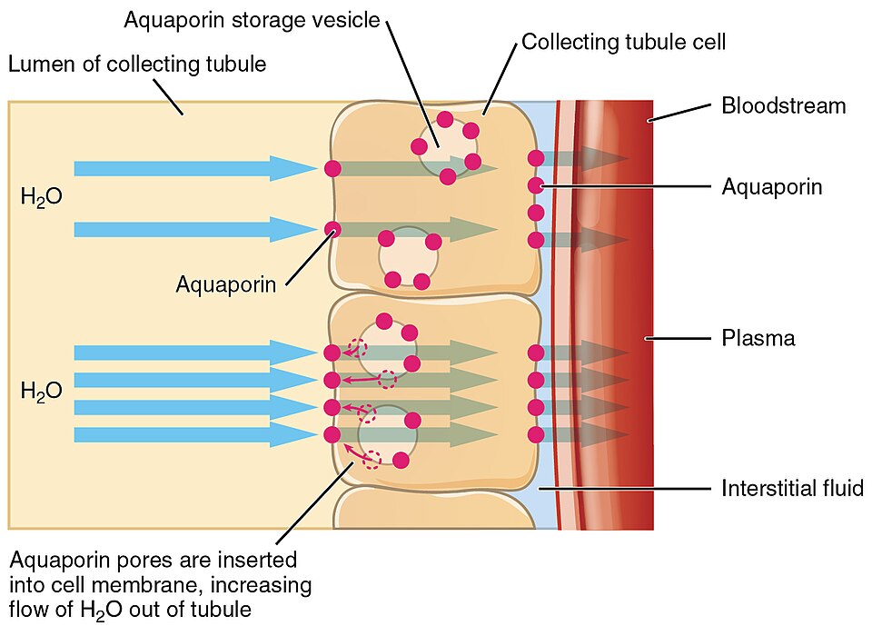
Aquaporins are channel proteins that allow water to cross membranes more easily. In osmoregulation, ADH increases the number of aquaporins in collecting duct membranes. This increases water reabsorption and helps restore blood water potential. Source
Blood glucose regulation: insulin and glucagon
· Blood glucose concentration is controlled by negative feedback.
· Insulin is released when blood glucose concentration is too high.
· Glucagon is released when blood glucose concentration is too low.
· Insulin acts on liver cells and muscle cells.
· Insulin causes cells to take up more glucose and stimulates conversion of glucose to glycogen by glycogenesis.
· Insulin lowers blood glucose concentration back to normal.
· Glucagon acts mainly on liver cells.
· Glucagon stimulates breakdown of glycogen to glucose by glycogenolysis.
· Glucagon raises blood glucose concentration back to normal.
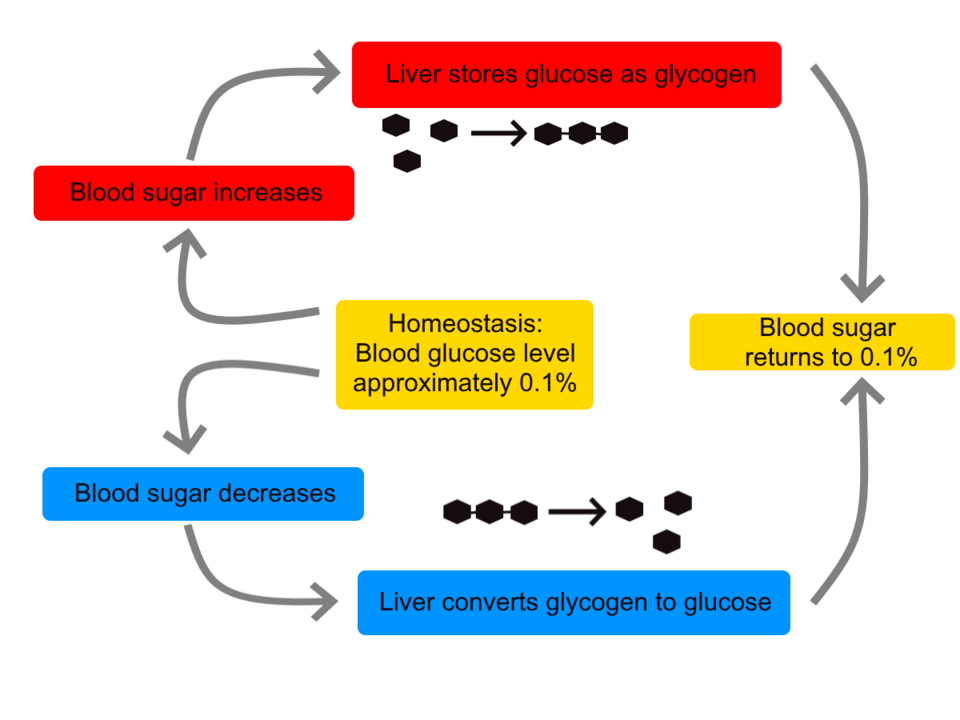
This diagram shows how insulin and glucagon have opposite effects on blood glucose concentration. Insulin lowers blood glucose by promoting glucose uptake and glycogen formation. Glucagon raises blood glucose by stimulating glycogen breakdown in the liver. Source
Cell signalling by glucagon
· Glucagon is a hormone that binds to a cell surface receptor on liver cells.
· Binding causes a conformational change in the receptor.
· This activates a G-protein.
· The G-protein stimulates adenylyl cyclase.
· Adenylyl cyclase converts ATP into cyclic AMP (cAMP).
· cAMP acts as a second messenger.
· cAMP activates protein kinase A.
· Protein kinase A starts an enzyme cascade by phosphorylation.
· The signal is amplified because each activated enzyme activates many more enzymes.
· Final response: enzyme activation causes glycogen breakdown, releasing glucose.
· Exam phrase: glucagon uses a second messenger pathway involving cAMP and an enzyme cascade.
Test strips and biosensors for glucose
· Test strips and biosensors measure glucose concentration in blood or urine.
· Both rely on enzyme specificity, especially glucose oxidase.
· Glucose oxidase catalyses oxidation of glucose, producing hydrogen peroxide.
· In test strips, peroxidase uses hydrogen peroxide to cause a colour change.
· The colour intensity is compared with a chart to estimate glucose concentration.
· In biosensors, the reaction produces an electrical signal.
· The size of the current is proportional to glucose concentration.
· Biosensors are more quantitative because they give a numerical reading.
· Exam phrase: glucose oxidase provides specificity because it acts only on glucose.
Common exam mistakes to avoid
· Do not say ADH is made by the posterior pituitary; it is released from the posterior pituitary.
· Do not say proteins are filtered into the nephron; large plasma proteins remain in the blood.
· Do not confuse ultrafiltration with selective reabsorption.
· Do not say insulin converts glucose directly; insulin stimulates cells to carry out glycogenesis.
· Do not forget that glucagon acts mainly on liver cells, not muscle cells.
· Do not describe positive feedback; blood glucose and osmoregulation use negative feedback.
Checklist: can you do this?
· Define homeostasis and explain why it is important in mammals.
· Explain a negative feedback pathway using stimulus → receptor → coordination → effector → response.
· Identify kidney and nephron structures from diagrams, photomicrographs and electron micrographs.
· Describe ultrafiltration, selective reabsorption and ADH-controlled osmoregulation.
· Explain blood glucose control, including insulin, glucagon, cAMP, enzyme cascades, test strips and biosensors.