Endocrine system
· Endocrine system = system of glands that secrete hormones directly into the blood.
· Hormones are chemical messengers that travel in blood to target cells with specific receptors.
· Endocrine responses are usually slower, longer-lasting and often affect many organs.
· Key syllabus examples: ADH, insulin and glucagon.
· ADH controls blood water potential by increasing water reabsorption in the collecting ducts of the kidney.
· Insulin lowers blood glucose by increasing glucose uptake by muscle/liver cells and promoting glycogenesis.
· Glucagon raises blood glucose by stimulating glycogen breakdown in liver cells.
Nervous system vs endocrine system
· Nervous system: impulses travel along neurones; uses electrical impulses and neurotransmitters.
· Endocrine system: hormones travel in blood plasma; uses chemical messengers only.
· Nervous responses are rapid, short-lived and localised.
· Endocrine responses are slower, longer-lasting and often widespread.
· Nervous effectors = muscles and glands; endocrine target cells = cells with specific hormone receptors.
· Exam comparison phrases: speed, duration, route of transmission, specificity, type of signal.
Sensory, motor and intermediate neurones
· Sensory neurone: transmits impulses from receptor cells to the CNS.
· Motor neurone: transmits impulses from the CNS to an effector, usually a muscle or gland.
· Intermediate neurones connect sensory neurones to motor neurones inside the CNS.
· A neurone has a cell body, dendrites, an axon and axon terminals.
· Myelin sheath insulates the axon and speeds up impulse transmission.
· Nodes of Ranvier are gaps in myelin where depolarisation occurs during saltatory conduction.
· Direction of impulse transmission is controlled by synapses and the refractory period.
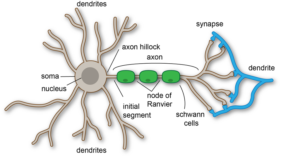
This diagram shows the basic structure of a neurone and the direction of information flow from dendrites to axon terminals. It is useful for comparing sensory and motor neurones and for linking structure to impulse transmission. Source
Sensory receptor cells and taste buds
· Sensory receptor cells detect a specific stimulus and convert it into a nervous impulse.
· A chemoreceptor detects chemicals, such as dissolved molecules in food.
· In a human taste bud, taste molecules bind to receptors on a chemoreceptor cell membrane.
· Binding causes ion channels to open or close, changing the membrane potential.
· If the change reaches threshold, an action potential is generated in the sensory neurone.
· The impulse then travels to the CNS for interpretation as taste.
· Key exam phrase: receptors act as transducers, converting stimulus energy into electrical signals.
Resting potential
· Resting potential = the potential difference across a neurone membrane when it is not transmitting an impulse.
· Inside of the axon is usually about –70 mV relative to the outside.
· Maintained by the sodium-potassium pump actively transporting 3 Na⁺ out and 2 K⁺ in.
· Membrane is more permeable to K⁺ than Na⁺, so K⁺ diffuses out through potassium channels.
· This makes the inside of the neurone negative compared with the outside.
· Resting potential requires ATP for active transport by the sodium-potassium pump.
Action potential
· Action potential = rapid change in membrane potential when a neurone transmits an impulse.
· Threshold must be reached for voltage-gated ion channels to open.
· Depolarisation: voltage-gated Na⁺ channels open, Na⁺ diffuses into the axon, inside becomes less negative.
· Membrane potential may reach about +30 mV.
· Repolarisation: Na⁺ channels close and voltage-gated K⁺ channels open, so K⁺ diffuses out.
· Hyperpolarisation may occur when too much K⁺ leaves.
· The resting potential is restored by closure of K⁺ channels and the sodium-potassium pump.
· Action potentials are all-or-nothing: a stronger stimulus increases frequency, not size, of impulses.
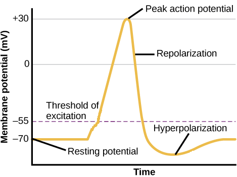
This diagram helps students link ion movement to changes in membrane potential during an action potential. It is especially useful for remembering the sequence Na⁺ influx → depolarisation → K⁺ efflux → repolarisation. Source
Refractory period
· Refractory period = time after an action potential when a neurone cannot immediately produce another action potential.
· Occurs because voltage-gated Na⁺ channels are inactivated and the membrane is recovering.
· Ensures impulses travel in one direction along the axon.
· Limits the maximum frequency of impulses.
· Higher stimulus intensity produces a higher frequency of action potentials, up to the limit set by the refractory period.
Saltatory conduction in myelinated neurones
· Myelinated neurones transmit impulses faster than unmyelinated neurones.
· Myelin sheath acts as an electrical insulator, preventing ion movement across covered regions of axon membrane.
· Depolarisation occurs only at nodes of Ranvier, where voltage-gated Na⁺ channels are concentrated.
· The impulse appears to “jump” from node to node = saltatory conduction.
· This increases speed and reduces energy use because fewer ions cross the membrane.
· Key exam phrase: local circuits carry depolarisation between adjacent nodes.
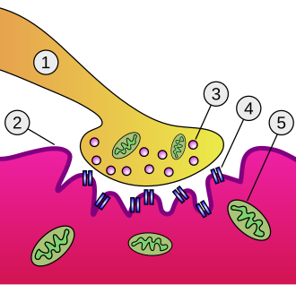
Cholinergic synapse
· Cholinergic synapse = synapse using acetylcholine (ACh) as the neurotransmitter.
· Structure includes presynaptic neurone, synaptic vesicles, synaptic cleft, postsynaptic membrane and receptor proteins.
· Action potential arrives at presynaptic knob, opening voltage-gated Ca²⁺ channels.
· Ca²⁺ ions enter the presynaptic knob and cause synaptic vesicles to fuse with the presynaptic membrane.
· ACh is released by exocytosis into the synaptic cleft.
· ACh diffuses across the cleft and binds to receptors on the postsynaptic membrane.
· Ligand-gated Na⁺ channels open, Na⁺ enters and depolarises the postsynaptic membrane.
· If threshold is reached, a new action potential is generated.
· Acetylcholinesterase breaks down ACh to stop continuous stimulation.
· Synapses ensure one-way transmission because vesicles are only presynaptic and receptors are only postsynaptic.
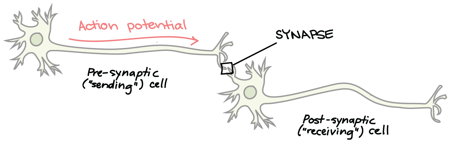
This resource shows how a nerve impulse is converted into a chemical signal at a synapse. It supports the CIE requirement to explain cholinergic synapse function, especially vesicle fusion, neurotransmitter diffusion and receptor binding. Source
Neuromuscular junction
· Neuromuscular junction = specialised cholinergic synapse between a motor neurone and striated muscle fibre.
· Motor neurone releases acetylcholine into the synaptic cleft.
· ACh binds to receptors on the sarcolemma, causing depolarisation.
· Depolarisation spreads along the sarcolemma and into the fibre through the T-tubule system.
· T-tubules carry the impulse deep into the muscle fibre.
· The impulse causes the sarcoplasmic reticulum to release Ca²⁺ ions.
· Ca²⁺ ions trigger interaction between actin and myosin, causing contraction.
· Acetylcholinesterase breaks down ACh so the muscle is not continuously stimulated.
This diagram shows the arrangement of structures at the neuromuscular junction. It is useful for linking synaptic transmission to stimulation of a muscle fibre. Source
Striated muscle ultrastructure
· Striated muscle contains parallel myofibrils made of repeating units called sarcomeres.
· Sarcomere = functional contractile unit between two Z-lines.
· Actin = thin filament attached to Z-lines.
· Myosin = thick filament with heads that form cross-bridges with actin.
· A band = region containing myosin; stays the same length during contraction.
· I band = region with actin only; becomes shorter during contraction.
· H zone = region with myosin only; becomes shorter during contraction.
· Z-lines move closer together during contraction.
· In electron micrographs, striations are due to regular arrangement of actin and myosin filaments.
Sliding filament model
· Sliding filament model: actin filaments slide past myosin filaments, shortening the sarcomere.
· Ca²⁺ ions bind to troponin, changing its shape.
· This moves tropomyosin away from myosin-binding sites on actin.
· Myosin heads bind to exposed sites on actin, forming cross-bridges.
· Myosin heads bend in a power stroke, pulling actin towards the centre of the sarcomere.
· ATP binds to myosin, causing myosin to detach from actin.
· ATP is hydrolysed to ADP + Pi, which re-cocks the myosin head.
· The cycle repeats while Ca²⁺ ions and ATP are available.
· When stimulation stops, Ca²⁺ ions are pumped back into the sarcoplasmic reticulum, tropomyosin blocks binding sites and the muscle relaxes.
· Essential roles: Ca²⁺ exposes binding sites, ATP detaches and reactivates myosin heads.
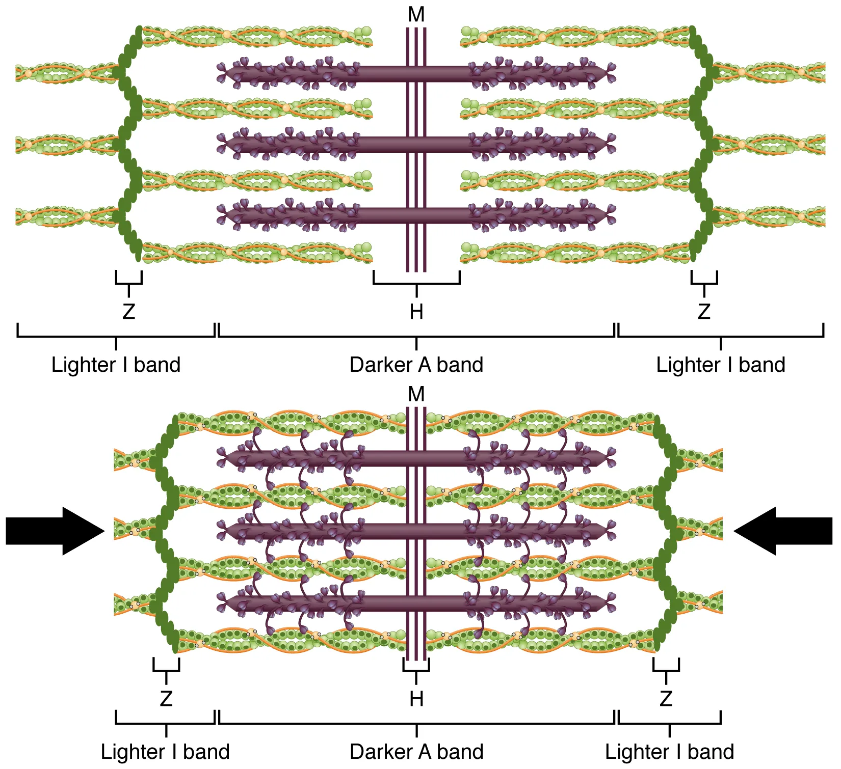
These diagrams show how sarcomeres shorten as thin filaments slide past thick filaments. They are ideal for revising the roles of actin, myosin, Ca²⁺, troponin, tropomyosin and ATP in contraction. Source
Common exam command words and answers
· Describe a neurone: name structures and state direction of impulse transmission.
· Explain an action potential: link ion channel opening to membrane potential changes.
· Compare nervous and endocrine systems: use clear paired contrasts such as fast/slow and short-lived/long-lasting.
· Explain synaptic transmission: include Ca²⁺ influx, vesicle fusion, ACh diffusion, receptor binding and acetylcholinesterase.
· Explain muscle contraction: include Ca²⁺ binding to troponin, tropomyosin movement, cross-bridge formation, ATP and sliding filaments.
· Interpret diagrams/electron micrographs: identify sarcomere, Z-line, A band, I band, H zone, actin and myosin.
Checklist: can you do this?
· Compare the nervous system and endocrine system using speed, duration, pathway and specificity.
· Explain resting potential, action potential, refractory period and saltatory conduction.
· Describe how a chemoreceptor in a taste bud can generate an impulse in a sensory neurone.
· Explain transmission across a cholinergic synapse and a neuromuscular junction.
· Use diagrams/electron micrographs to explain sarcomere structure and the sliding filament model.