Fluid mosaic model
· Fluid mosaic model = membranes are a fluid phospholipid bilayer with a mosaic of proteins and other molecules embedded or attached.
· Fluid = phospholipids and some proteins can move laterally within the bilayer.
· Mosaic = many different components, including phospholipids, cholesterol, proteins, glycolipids and glycoproteins.
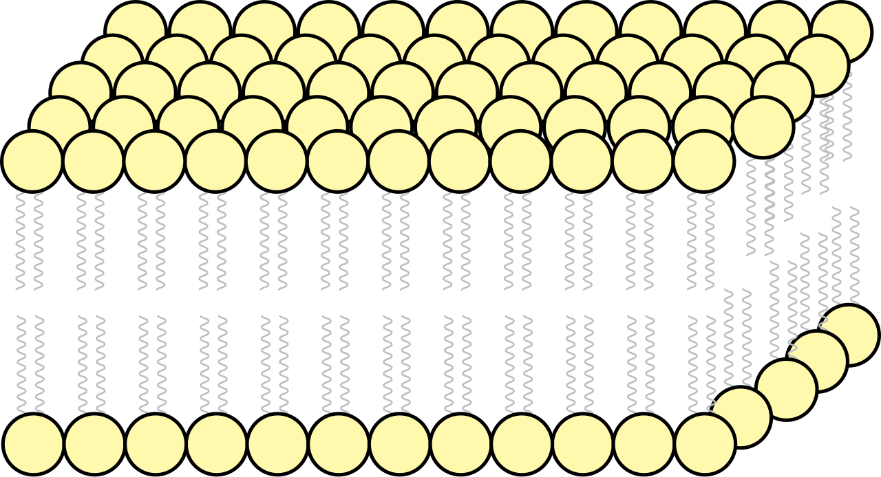
· Phospholipids form a bilayer because their hydrophilic phosphate heads face water and their hydrophobic fatty acid tails face inwards, away from water.
· The bilayer forms due to hydrophobic interactions between fatty acid tails and hydrophilic interactions between phosphate heads and water.
· Membrane proteins are arranged according to interactions between their hydrophobic regions and the hydrophobic core of the bilayer.
This diagram shows the fluid mosaic model, with molecules arranged in a flexible membrane structure. It is useful for identifying the bilayer and embedded membrane components. Source
Phospholipids
· Phospholipids are the main structural component of cell surface membranes.
· Each phospholipid has a hydrophilic head and two hydrophobic tails.
· The hydrophilic heads face the aqueous extracellular fluid and cytoplasm.
· The hydrophobic tails face inwards, forming a hydrophobic core.
· The hydrophobic core makes the membrane partially permeable by restricting many polar molecules and ions.
· Small non-polar molecules can pass through the phospholipid bilayer more easily than charged or polar substances.
Cholesterol
· Cholesterol is found between phospholipid molecules in cell surface membranes.
· It increases membrane stability by helping phospholipids pack together.
· It regulates membrane fluidity, preventing the membrane from becoming too fluid or too rigid.
· It reduces membrane permeability to some small polar molecules and ions.
· Exam phrase: cholesterol maintains stability and fluidity of the membrane.
Membrane proteins
· Channel proteins form hydrophilic pores that allow specific ions or polar molecules to move across the membrane.
· Carrier proteins bind specific substances and change shape to move them across the membrane.
· Transport proteins are important for facilitated diffusion and active transport.
· Cell surface receptors are proteins that bind specific signalling molecules called ligands.
· Some proteins act as enzymes or help attach the membrane to internal cell structures.
· Proteins may be intrinsic/integral if embedded in the bilayer or extrinsic/peripheral if attached to the membrane surface.
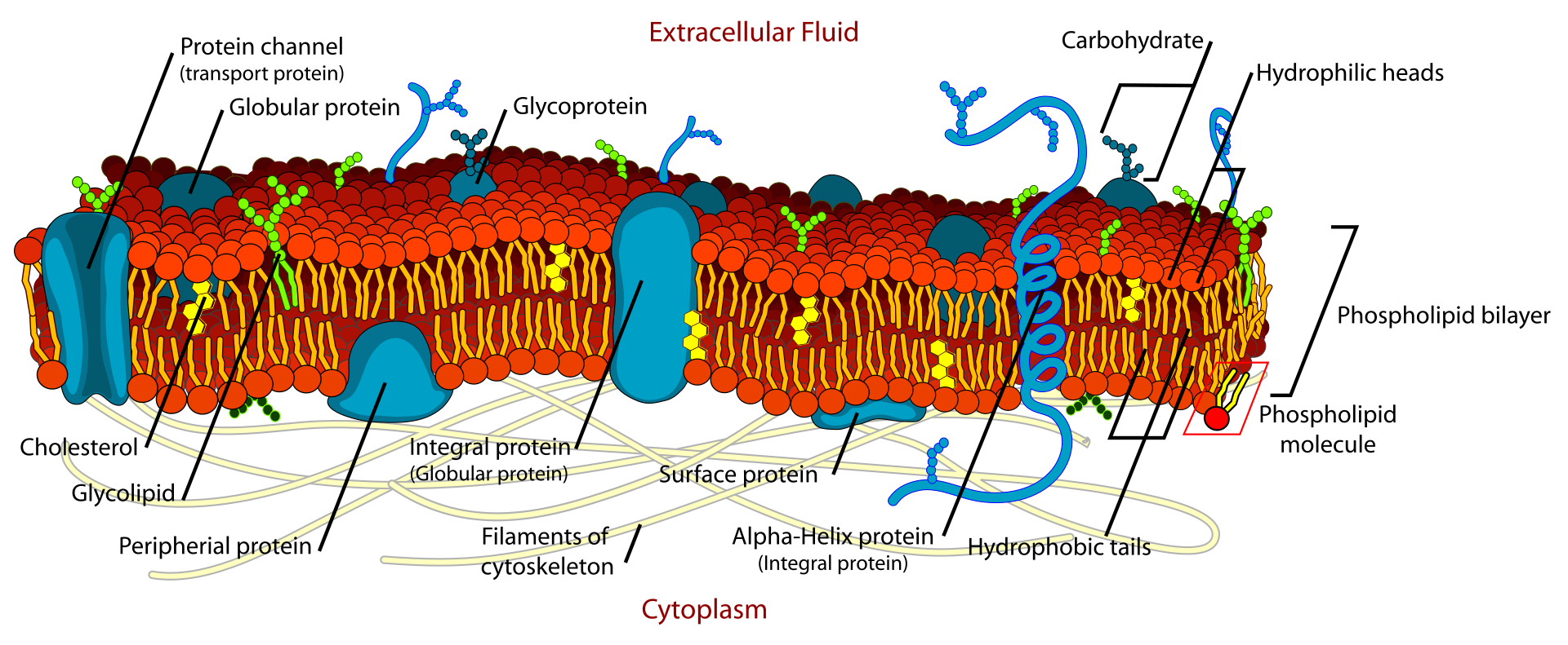
This labelled diagram shows how proteins, cholesterol, glycoproteins and glycolipids are arranged in the membrane. It is useful for revising both the structure and roles of membrane components. Source
Glycolipids and glycoproteins
· Glycolipids = lipids with attached carbohydrate chains.
· Glycoproteins = proteins with attached carbohydrate chains.
· Carbohydrate chains project from the outer surface of the cell surface membrane.
· Glycolipids and glycoproteins are involved in cell recognition and cell signalling.
· They can act as cell surface antigens, allowing cells to be recognised as self or non-self.
· Exam link: cell surface antigens are important in immune recognition.
Roles of membrane components
· Phospholipids: form the bilayer, create a hydrophobic barrier, allow membrane fluidity.
· Cholesterol: improves stability, controls fluidity, reduces permeability.
· Channel proteins: allow specific ions or polar molecules through by facilitated diffusion.
· Carrier proteins: transport specific molecules by changing shape; used in facilitated diffusion and active transport.
· Receptor proteins: bind ligands and trigger cell signalling.
· Glycoproteins/glycolipids: involved in cell recognition, cell adhesion, cell signalling and cell surface antigens.
Cell signalling
· Cell signalling allows cells to communicate and produce specific responses.
· Stage 1: cells secrete specific chemicals called ligands.
· Stage 2: ligands are transported to target cells.
· Stage 3: ligands bind to specific cell surface receptors on target cells.
· Binding is specific because receptor shape is complementary to the ligand.
· Ligand binding can cause a conformational change in the receptor, leading to a response inside the target cell.
· Exam phrase: only target cells with the correct receptor respond to the ligand.
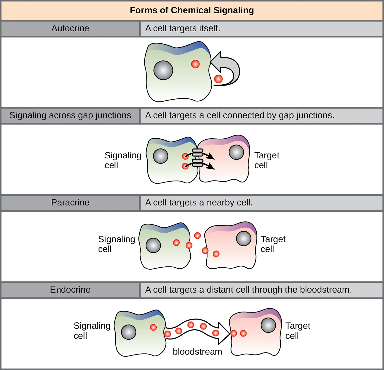
This image supports the idea that ligands bind to specific receptors on target cells. The binding event begins a signalling process that produces a specific cellular response. Source
Exam command words and common mark points
· Describe membrane structure: mention phospholipid bilayer, hydrophilic heads, hydrophobic tails, proteins, cholesterol, glycolipids and glycoproteins.
· Explain bilayer formation: refer to hydrophobic interactions between tails and hydrophilic interactions between heads and water.
· Explain permeability: refer to the hydrophobic core restricting ions and many polar molecules.
· Explain transport roles: distinguish channel proteins from carrier proteins.
· Explain recognition/signalling: refer to glycoproteins, glycolipids, cell surface antigens, ligands and receptors.
Checklist: can you do this?
· Describe the fluid mosaic model using hydrophilic and hydrophobic interactions.
· Identify and state the arrangement of cholesterol, glycolipids and glycoproteins in membranes.
· Explain the roles of phospholipids, cholesterol, proteins, glycolipids and glycoproteins.
· Compare the roles of channel proteins and carrier proteins in transport.
· Outline the stages of cell signalling: ligand secretion, ligand transport, receptor binding and response.