Chromosome Behaviour in Mitosis
· Mitosis = division of the nucleus to produce two genetically identical nuclei.
· Occurs after DNA replication in S phase, so each chromosome has two sister chromatids joined at a centromere.
· Key sequence: prophase → metaphase → anaphase → telophase.
· Cytokinesis follows mitosis and divides the cytoplasm, producing two cells.
· In exams, focus on what chromosomes look like, where they are, and what the spindle / nuclear envelope / cell membrane is doing.
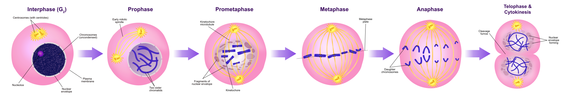
This diagram summarises the main stages of mitosis, showing how duplicated chromosomes are arranged and separated. Use it to practise identifying prophase, metaphase, anaphase and telophase from chromosome position and spindle behaviour. Source
Prophase
· Chromosomes condense: long chromatin coils tightly and becomes visible as distinct chromosomes.
· Each chromosome consists of two sister chromatids joined by a centromere.
· Nucleolus disappears.
· Nuclear envelope breaks down, allowing spindle fibres to access chromosomes.
· Spindle forms from microtubules.
· In animal cells, spindle fibres organise from centrioles/centrosomes moving to opposite poles.
· In plant cells, spindle forms without centrioles in most higher plants.
· Exam clue: chromosomes look short, thick and scattered, not yet aligned.
Metaphase
· Chromosomes attach to spindle fibres at their centromeres.
· Chromosomes line up along the equator / metaphase plate of the cell.
· Sister chromatids are still joined at the centromere.
· Spindle fibres extend from opposite poles and attach to opposite sides of each chromosome.
· Exam clue: chromosomes are arranged in a single line across the centre of the cell.
· Metaphase ensures each daughter nucleus receives one copy of each chromosome.
Anaphase
· Centromeres divide.
· Sister chromatids separate and are pulled to opposite poles of the cell.
· Once separated, each chromatid is considered a daughter chromosome.
· Movement occurs because spindle fibres shorten.
· The cell begins to elongate as chromosome sets move apart.
· Exam clue: chromosomes often appear V-shaped or pulled apart toward opposite poles.
· Anaphase ensures both new nuclei receive identical sets of chromosomes.
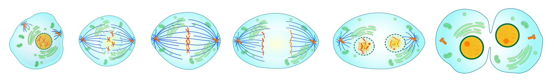
This sequence is useful for seeing the transition from metaphase to anaphase. Focus on how sister chromatids separate and move toward opposite poles, forming identical chromosome sets. Source
Telophase
· Chromosomes reach opposite poles of the cell.
· Chromosomes decondense and become less visible.
· Nuclear envelopes reform around each set of chromosomes.
· Nucleoli reappear.
· Spindle breaks down.
· Telophase produces two genetically identical nuclei.
· Exam clue: two new nuclei are visible, often with chromosomes becoming harder to distinguish.
Cytokinesis and Cell Surface Membrane Behaviour
· Cytokinesis = division of the cytoplasm, not the nucleus.
· In animal cells, the cell surface membrane constricts to form a cleavage furrow.
· The cleavage furrow deepens until the cell separates into two daughter cells.
· In plant cells, vesicles form a cell plate across the equator.
· The cell plate develops into a new cell wall between daughter cells.
· Both daughter cells contain genetically identical nuclei produced by mitosis.
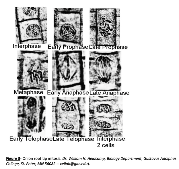
This image is useful for recognising mitosis in plant cells, especially in an onion root tip. It also shows why plant cytokinesis differs from animal cytokinesis: plant cells form a cell plate rather than a cleavage furrow. Source
Interpreting Microscope Slides and Photomicrographs
· Identify stages by chromosome position, not by guessing from cell shape.
· Interphase: nucleus visible; chromosomes not clearly visible as separate structures.
· Prophase: chromosomes visible and condensed; not aligned.
· Metaphase: chromosomes aligned at the equator.
· Anaphase: sister chromatids moving to opposite poles.
· Telophase: two nuclei forming; chromosomes decondensing.
· In root tip slides, many cells may be in interphase because it is the longest part of the cell cycle.
· For drawings, use clear outlines, no shading, and label key features such as chromosomes, spindle fibres, nuclear envelope and cell plate if visible.
Common Exam Mistakes
· Do not say mitosis produces four cells; that is meiosis.
· Do not say chromosomes “disappear” in telophase; they decondense and become less visible.
· Do not confuse chromosomes with chromatids: chromatids separate in anaphase.
· Do not say cytokinesis is part of nuclear division; it is division of the cytoplasm.
· Do not describe crossing over or homologous chromosome pairing; these occur in meiosis, not mitosis.
· Do not ignore plant/animal differences in cytokinesis.
Checklist: can you do this?
· Describe chromosome behaviour in prophase, metaphase, anaphase and telophase.
· Explain what happens to the nuclear envelope, spindle and cell surface membrane during mitosis/cytokinesis.
· Identify mitotic stages from photomicrographs, diagrams and microscope slides.
· Distinguish sister chromatids, chromosomes, centromeres and daughter chromosomes.
· Compare cytokinesis in plant cells and animal cells.