Structure of Transport Tissues
· Transport tissues in flowering plants are xylem and phloem.
· They occur together in vascular bundles in stems and leaves, and in the central vascular cylinder / stele of roots.
· Required plant type: herbaceous dicotyledonous plants.
· In exams, focus on distribution, drawing accuracy, labelling, and structure-to-function links.
· Do not confuse: xylem transports water and mineral ions; phloem transports assimilates such as sucrose and amino acids.
Distribution of xylem and phloem in stems
· In a dicot stem transverse section, vascular bundles are arranged in a ring near the edge of the stem.
· In each vascular bundle, xylem is found on the inner side, closer to the pith.
· Phloem is found on the outer side, closer to the cortex / epidermis.
· Cambium may be shown between xylem and phloem, but focus for 7.1 is mainly xylem and phloem position.
· Plan diagrams should show tissue outlines only, not individual cells.
· Use single, clear, unbroken lines and label epidermis, cortex, vascular bundles, xylem, phloem and pith if visible.
This diagram shows the relative positions of xylem and phloem in a stem vascular bundle. It is useful for remembering that xylem is towards the inside and phloem is towards the outside in a dicot stem. Source
Distribution of xylem and phloem in roots
· In a dicot root transverse section, vascular tissue is in the centre.
· Xylem often forms a central star / X-shaped structure.
· Phloem is found in groups between the arms of the xylem.
· The cortex forms a large region around the central vascular tissue.
· The endodermis surrounds the central vascular tissue, but detailed transport mechanisms are topic 7.2.
· Plan diagrams should show the central xylem shape, phloem between xylem arms, and the surrounding cortex clearly.
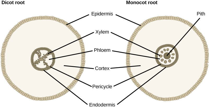
This image shows the typical distribution of xylem and phloem in a dicot root. The key exam point is that xylem is central and phloem lies between the arms of the xylem. Source
Distribution of xylem and phloem in leaves
· In a dicot leaf transverse section, xylem and phloem are found in the vascular bundle / vein.
· Xylem is usually on the upper side of the vascular bundle, nearer the upper epidermis.
· Phloem is usually on the lower side of the vascular bundle, nearer the lower epidermis.
· The vascular bundle is usually surrounded by supporting tissue and lies within the mesophyll.
· In plan diagrams, show the upper epidermis, palisade mesophyll, spongy mesophyll, vascular bundle, xylem, phloem and lower epidermis if visible.
· Do not draw every cell in a plan diagram; show proportions and tissue boundaries.
This image shows where transport tissues are found inside a leaf vein. It helps students remember that in a leaf vascular bundle, xylem is uppermost and phloem is below it. Source
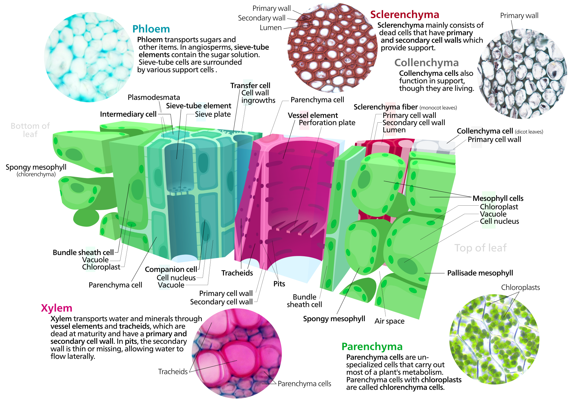
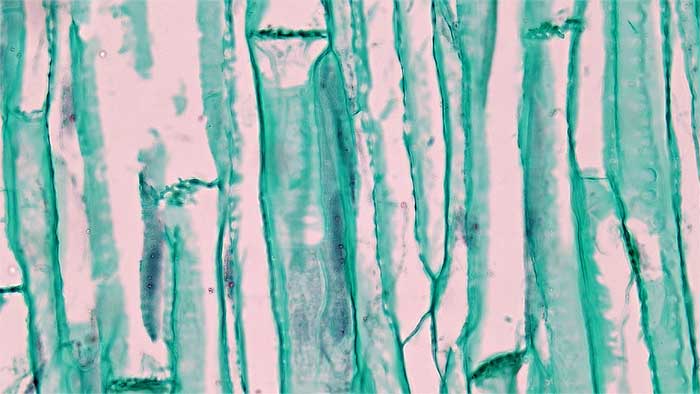
Xylem vessel elements: structure and function
· Xylem vessel elements are long cells joined end-to-end to form continuous tubes.
· They are dead at maturity, so the lumen is empty and water can move through with little resistance.
· End walls break down or are absent, forming an uninterrupted pathway for water movement.
· Lignin is deposited in the cell walls, making them thick, strong and waterproof.
· Lignified walls prevent collapse when xylem is under tension during water transport.
· Pits in the walls allow water to move sideways between vessels and surrounding tissues.
· Function: transport water and mineral ions from roots to stems and leaves, and provide mechanical support.
· When drawing from micrographs, label lumen, lignified wall, pits and end wall absent / perforation if visible.
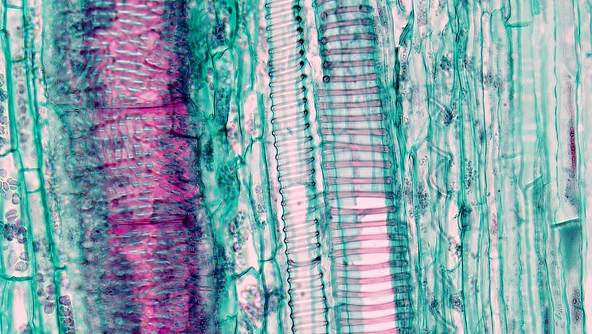
This photomicrograph shows xylem elements from a herbaceous dicot stem. It is useful for practising recognition of thickened xylem walls and drawing labelled xylem vessel elements from microscope images. Source
Phloem sieve tube elements: structure and function
· Phloem sieve tube elements are living cells arranged end-to-end to form sieve tubes.
· They transport assimilates, especially sucrose and amino acids.
· They have sieve plates between cells, containing pores that allow phloem sap to pass through.
· They have very little cytoplasm and few organelles, reducing resistance to flow.
· They lack a nucleus at maturity, so they depend on companion cells for metabolic support.
· Function: transport organic substances from sources to sinks.
· When drawing from micrographs, label sieve tube element, sieve plate, pore and companion cell if visible.
Companion cells: structure and function
· Companion cells are closely associated with sieve tube elements.
· They contain a nucleus, dense cytoplasm and many mitochondria.
· They are metabolically active and help maintain the living sieve tube elements.
· Many mitochondria provide ATP for active processes involved in phloem transport.
· Plasmodesmata connect companion cells to sieve tube elements, allowing movement of substances between them.
· Function: support sieve tube elements and help load/unload assimilates.
· Structure-function link: many mitochondria + close contact with sieve tubes = efficient support for phloem transport.
This image shows the close association between sieve tube elements and companion cells. It supports the key exam link that sieve tube elements rely on companion cells because they have reduced contents and no nucleus at maturity. Source
Drawing skills for 7.1
· For plan diagrams, draw tissues, not individual cells.
· Use large diagrams, covering at least half the available space.
· Use clear outlines, no sketching, shading or colouring.
· Keep proportions realistic, especially the relative positions of xylem and phloem.
· For cell drawings, draw individual structures from microscope slides, photomicrographs or electron micrographs.
· Label only what is visible: xylem vessel element, lignified wall, lumen, pit, sieve tube element, sieve plate, companion cell.
· Use a ruler for label lines and make sure label lines do not cross.
· Do not invent structures that cannot be seen in the image.
Common exam comparisons
· Xylem: dead cells, hollow lumen, lignified walls, transports water and mineral ions, also provides support.
· Phloem: living tissue, includes sieve tube elements and companion cells, transports assimilates.
· Sieve tube elements: reduced contents, sieve plates, no nucleus at maturity, adapted for flow of phloem sap.
· Companion cells: many mitochondria, nucleus, dense cytoplasm, adapted for metabolic support of sieve tube elements.
· Stem distribution: vascular bundles in a ring, xylem inside, phloem outside.
· Root distribution: central star / X-shaped xylem, phloem between xylem arms.
· Leaf distribution: xylem usually upper, phloem usually lower in the vascular bundle.
Checklist: can you do this?
· Draw plan diagrams of dicot stem, root and leaf transverse sections from slides or photomicrographs.
· Describe the distribution of xylem and phloem in dicot stems, roots and leaves.
· Draw and label xylem vessel elements, sieve tube elements and companion cells from microscope images.
· Explain how xylem vessel structure is related to water transport and support.
· Explain how sieve tube elements and companion cells are adapted for phloem transport.