The gas exchange system
· Main role: gas exchange system takes in oxygen (O₂) and removes carbon dioxide (CO₂).
· Gas exchange occurs between air in the alveoli and blood in capillaries.
· Key structures: lungs, trachea, bronchi, bronchioles, alveoli, capillary network.
· Exam focus: know structure, tissue distribution, functions, and how to recognise structures in microscope images.
Structure of the human gas exchange system
· Lungs = organs containing branching airways and many alveoli for gas exchange.
· Trachea = main airway from throat to chest; supported by cartilage.
· Bronchi = two main branches from trachea, one entering each lung.
· Bronchioles = smaller branching airways leading towards alveoli.
· Alveoli = tiny air sacs with thin squamous epithelium and surrounding capillary network.
· Capillary network = brings deoxygenated blood close to alveolar air and carries oxygenated blood away.
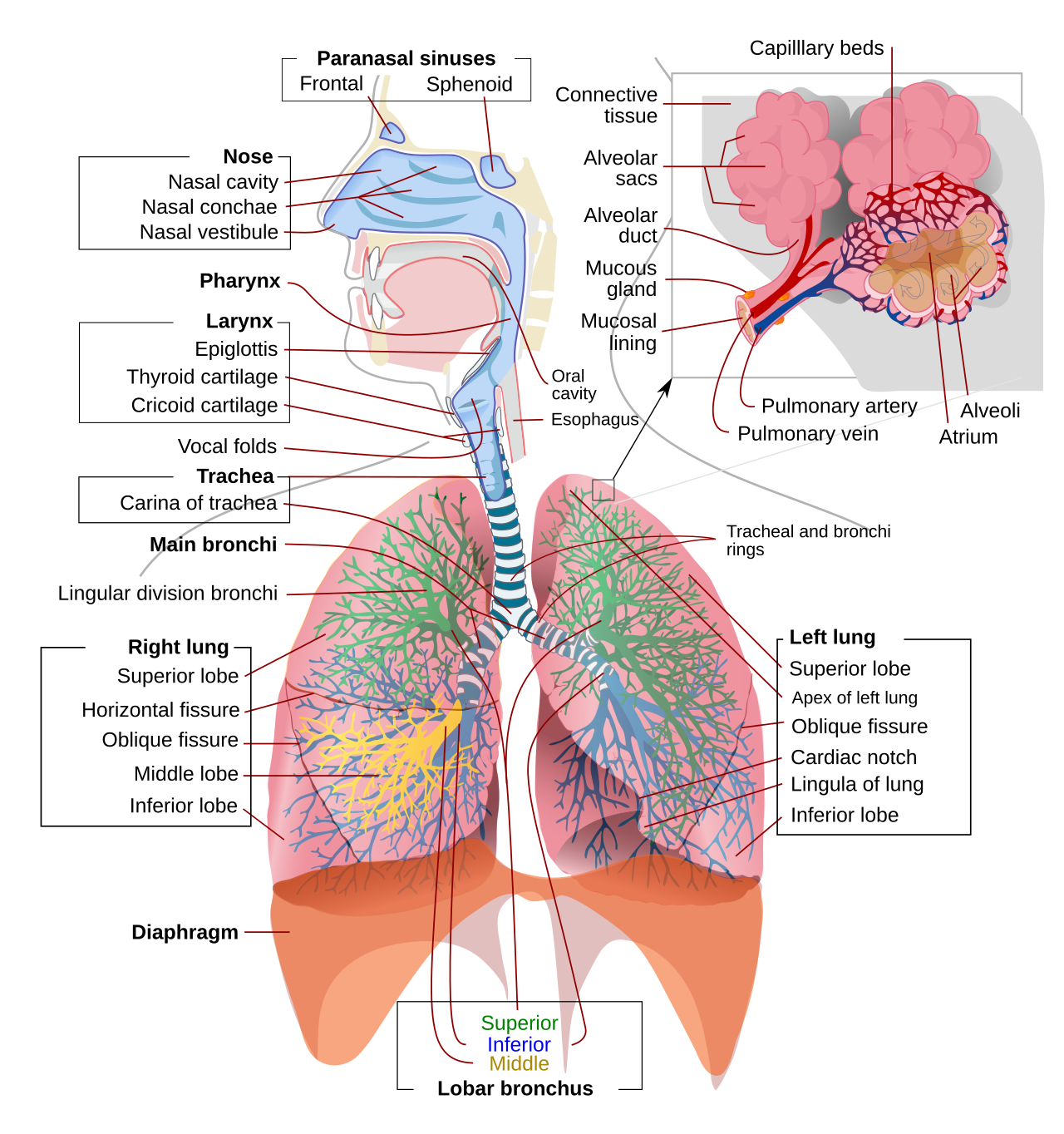
This diagram shows the pathway of air through the human gas exchange system. It is useful for learning the order of structures from trachea → bronchi → bronchioles → alveoli. Source
Distribution of tissues in the gas exchange system
· Cartilage: present in trachea and bronchi; absent from bronchioles and alveoli.
· Ciliated epithelium: lines trachea, bronchi and many bronchioles.
· Goblet cells: found in trachea and bronchi; produce mucus.
· Mucous glands: present in larger airways; secrete mucus to trap particles/pathogens.
· Smooth muscle: found in trachea, bronchi and especially bronchioles; controls airway diameter.
· Squamous epithelium of alveoli: very thin cells forming alveolar walls.
· Capillaries: closely surround alveoli, reducing diffusion distance for gas exchange.
This page helps students recognise ciliated epithelium, goblet cells and airway wall tissues. These are key features for identifying trachea and bronchi in microscope images. Source
Recognising structures in microscope images
· Trachea: large airway, thick wall, cartilage, ciliated epithelium, goblet cells, mucous glands.
· Bronchus: airway with cartilage plates, smooth muscle, ciliated epithelium and goblet cells.
· Bronchiole: smaller airway, no cartilage, usually more obvious smooth muscle relative to size.
· Alveoli: many small air spaces; very thin walls made of squamous epithelium.
· Capillaries: tiny blood vessels close to alveolar walls; may contain red blood cells.
· In plan diagrams of trachea and bronchus TS, show general tissue layers but do not draw individual cells in detail.
Maintaining health of the gas exchange system
· Goblet cells secrete mucus.
· Mucous glands also secrete mucus in larger airways.
· Mucus traps dust, pathogens and other particles before they reach alveoli.
· Ciliated epithelial cells move mucus upwards towards the throat.
· The mucus is then swallowed or removed, helping prevent infection and blockage of alveoli.
· Damage to cilia reduces mucus removal, increasing risk of infection and impaired gas exchange.
Functions of key tissues
· Cartilage: supports airways and prevents trachea and bronchi from collapsing.
· Smooth muscle: contracts or relaxes to change the diameter of airways, especially bronchioles.
· Elastic fibres: stretch during inhalation and recoil during exhalation, helping move air out.
· Squamous epithelium: forms thin alveolar walls, giving a short diffusion distance.
· Capillaries: maintain steep diffusion gradients by bringing blood close to alveolar air.
Gas exchange at the alveoli
· Oxygen diffuses from alveolar air into blood capillaries.
· Carbon dioxide diffuses from blood capillaries into alveolar air.
· Diffusion occurs down concentration gradients.
· Alveoli are adapted for efficient gas exchange by having:
· Large surface area from many alveoli.
· Short diffusion distance due to thin alveolar and capillary walls.
· Good blood supply from dense capillary network.
· Ventilation maintains oxygen and carbon dioxide gradients.
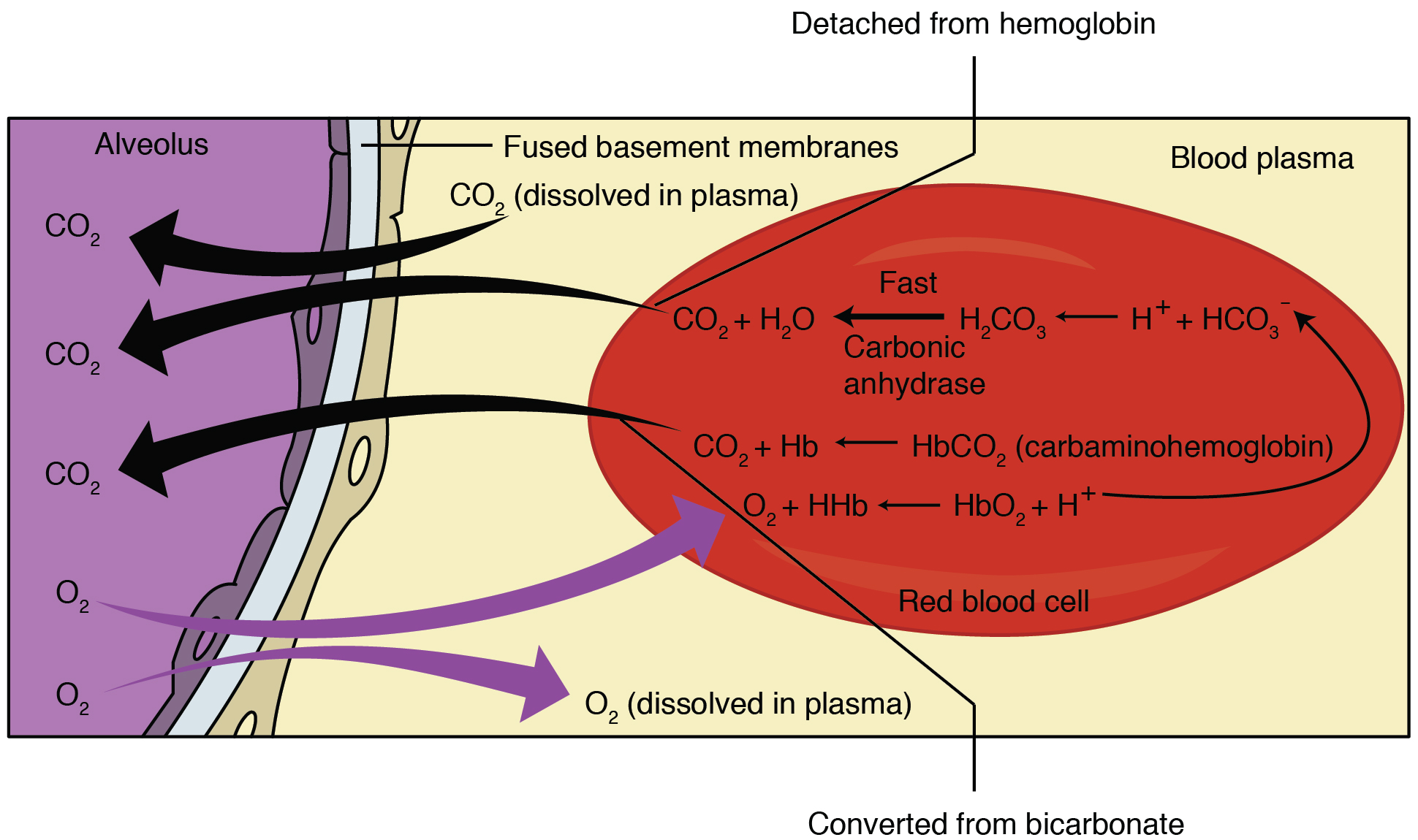
This diagram shows O₂ diffusing from alveolus to blood and CO₂ diffusing from blood to alveolus. It is ideal for revising the direction of gas movement and the role of the respiratory membrane. Source
Exam skills: plan diagrams and image interpretation
· For plan diagrams, draw tissue outlines only; avoid shading and unnecessary cell detail.
· Label key tissues such as cartilage, epithelium, smooth muscle, lumen and mucous glands where visible.
· Use relative size and wall structure to distinguish trachea, bronchus, bronchiole and alveoli.
· In photomicrographs, identify cartilage as supportive tissue in larger airways.
· Identify alveoli by their many air spaces and very thin walls.
· Link every visible structure to its function, e.g. squamous epithelium → short diffusion distance.
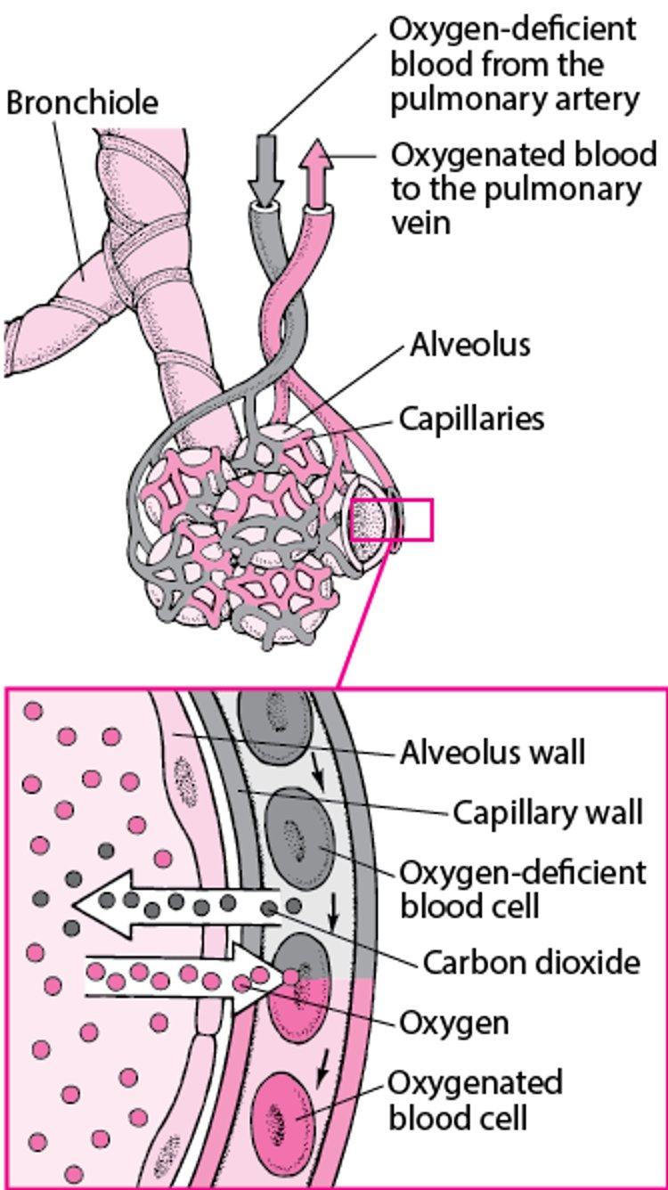
This diagram clearly shows how the alveoli and capillaries are arranged closely together. It reinforces why a short diffusion pathway and good blood supply make gas exchange efficient. Source
Common exam traps
· Do not say gas exchange occurs in the bronchi or trachea; it occurs mainly in the alveoli.
· Do not confuse bronchi with bronchioles: bronchi have cartilage, bronchioles do not.
· Do not describe alveolar walls as “thick”; they are made of thin squamous epithelium.
· Do not say cilia trap pathogens; mucus traps, while cilia move mucus.
· Always link structure to function: thin walls, large surface area, capillary network, ventilation.
Checklist: can you do this?
· Describe the structure of the lungs, trachea, bronchi, bronchioles, alveoli and capillary network.
· State where cartilage, ciliated epithelium, goblet cells, smooth muscle, squamous epithelium and capillaries are found.
· Recognise trachea, bronchi, bronchioles and alveoli in microscope images.
· Draw plan diagrams of transverse sections of trachea and bronchus walls.
· Explain gas exchange between alveolar air and capillary blood using diffusion gradients.