Structure and function of connective tissues and joints
· Syllabus focus: how connective tissue structure and joint structure enable movement while providing stability. Cellular-level detail is not assessed.
· Core exam idea: there is always a trade-off between mobility and stability: joints that allow more movement usually provide less stability, while highly stable joints usually allow less movement.
Connective tissues: structure → function
· Bone = hard supportive connective tissue; provides levers, shape, protection, and attachment points for muscles/tendons.
· Ligaments = strong bands connecting bone to bone; increase joint stability and limit excessive or abnormal movement.
· Cartilage = smooth, tough, flexible tissue; reduces friction, absorbs shock, and supports smoother movement at joint surfaces.
· Tendons = strong connective tissue connecting muscle to bone; transmit muscle force to bones to produce movement.
· Fascia = connective tissue sheets surrounding/supporting muscles and other structures; helps transmit force and maintain tissue organization.
· Exam link: connective tissues increase stability and permit movement, but each tissue contributes differently.
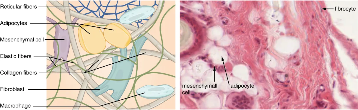
This image helps students compare the structural features of connective tissues involved in movement and stability. Focus on dense regular connective tissue for tendons/ligaments and supportive connective tissue for bone/cartilage. Source
Types of articulations
· Articulation = a joint, where bones or bone and cartilage meet.
· The three main structural types are fibrous, cartilaginous, and synovial joints.
· Fibrous joints = bones joined by fibrous connective tissue; usually very stable with little movement.
· Cartilaginous joints = bones joined by hyaline cartilage or fibrocartilage; allow limited movement with good stability.
· Synovial joints = bones meet inside a fluid-filled joint cavity; allow the greatest range of movement.
· Functional classification: joints range from immobile, to slightly mobile, to freely movable.
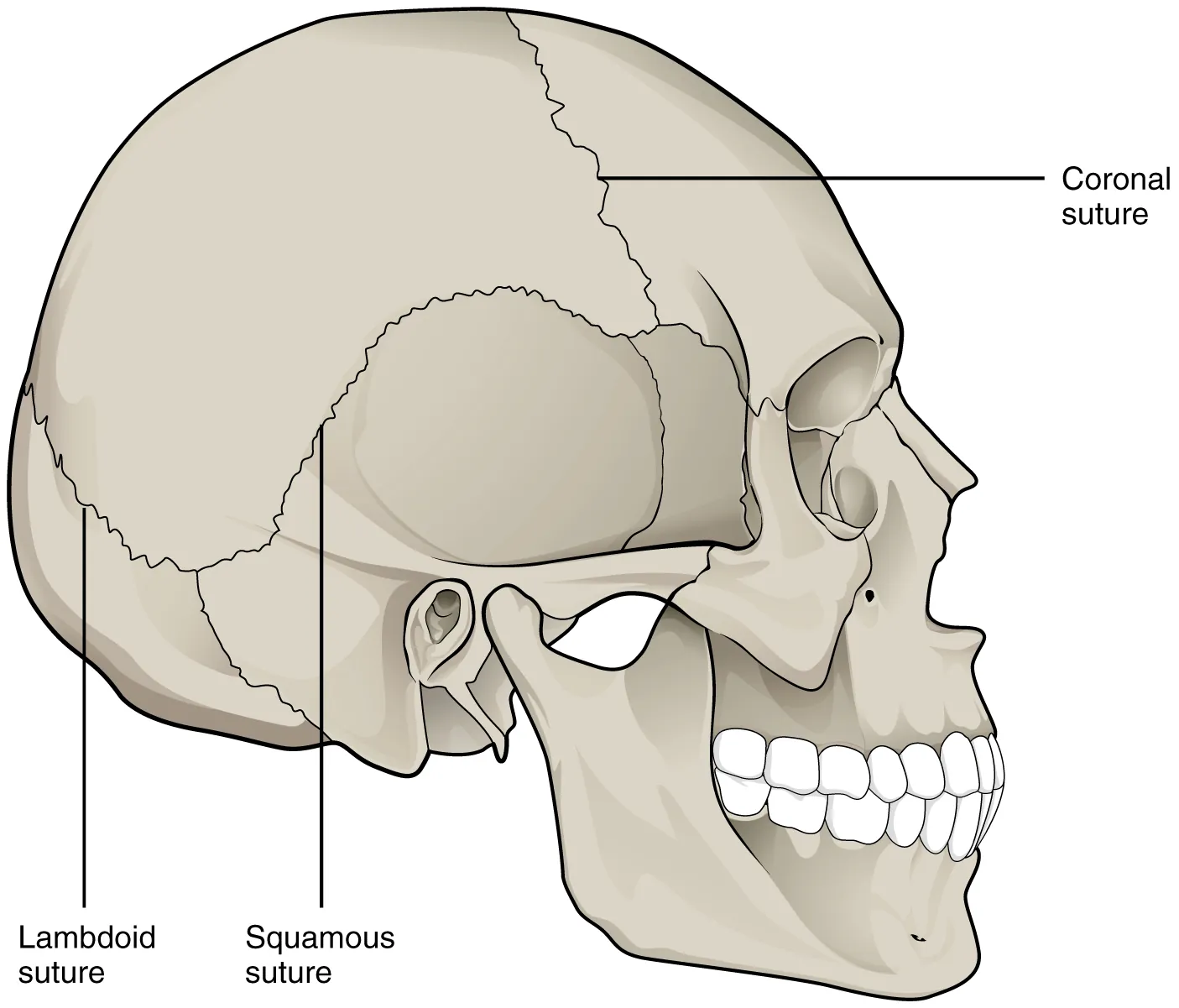
These images show how joint structure affects movement. Sutures are highly stable with almost no movement, while intervertebral discs allow limited movement between vertebrae. Source
Synovial joints: key structure
· Synovial joints are the most important for sport because they allow free movement.
· Key structures: joint cavity, synovial fluid, articular cartilage, articular capsule, and ligaments.
· Synovial fluid lubricates the joint and reduces friction.
· Articular cartilage covers bone ends, allowing smoother movement and reducing wear.
· Articular capsule surrounds the joint and helps enclose the joint cavity.
· Ligaments reinforce the joint and resist excessive movement.
· Tendons and muscles can provide extra dynamic stability by supporting the joint during movement.
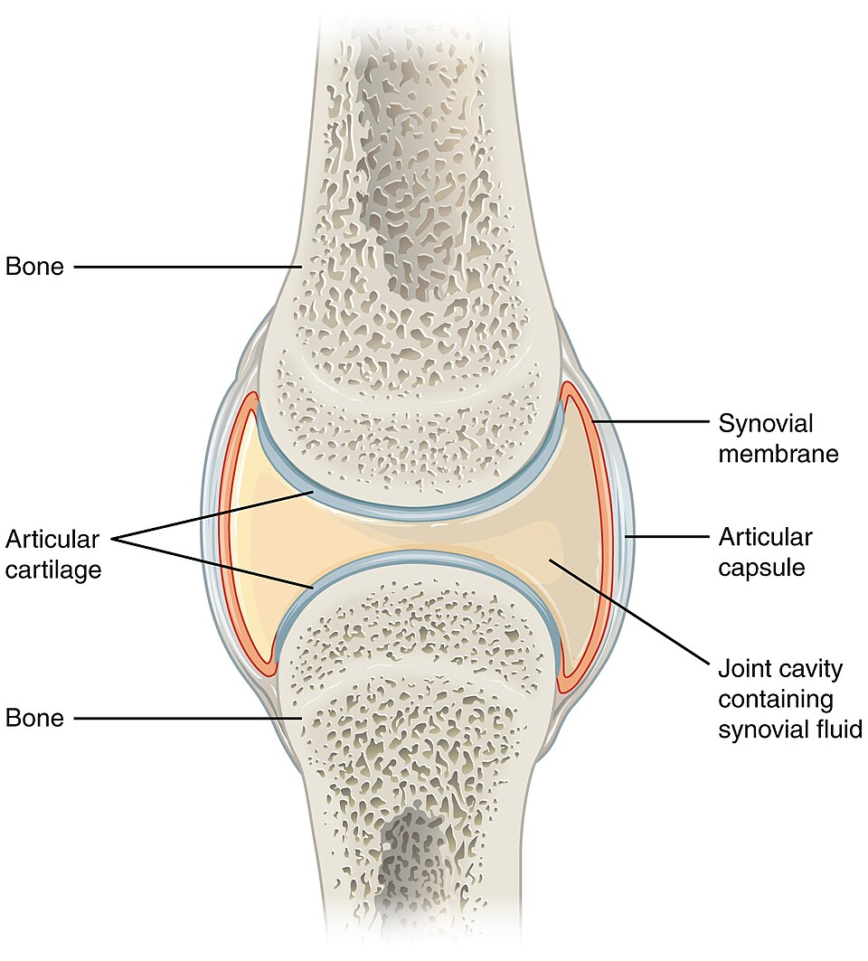
This diagram shows the main structures that make synovial joints highly mobile. It is especially useful for linking synovial fluid and articular cartilage to reduced friction and smooth movement. Source
Classes of synovial joints
· Plane/gliding joint = sliding movement; example: joints between some wrist/ankle bones.
· Hinge joint = movement mainly in one plane; example: elbow or knee.
· Pivot joint = rotation around one axis; example: atlas–axis joint in the neck.
· Condyloid joint = movement in two planes; example: wrist joint.
· Saddle joint = movement in two planes with strong control; example: thumb joint.
· Ball-and-socket joint = movement in multiple planes; example: shoulder or hip.
· Exam link: compare joints by range of movement, axes of movement, and stability.
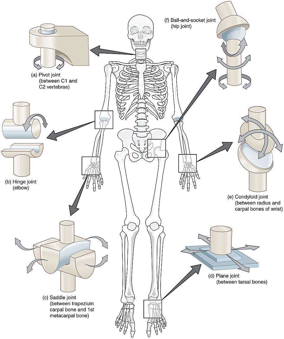
This image is ideal for memorising the main synovial joint classes. Use it to link each joint shape to the movement it permits and the stability it provides. Source
Stability vs movement in sport
· High stability, low movement: fibrous joints and many cartilaginous joints.
· High movement, lower stability: many synovial joints, especially the shoulder.
· Hip vs shoulder comparison: both are ball-and-socket joints, but the hip is generally more stable because strong ligaments restrict movement; the shoulder has greater range of motion but less inherent stability.
· Joint stability depends on joint shape, ligaments, tendons, muscles, cartilage, and surrounding fascia.
· Sporting relevance: athletes need enough mobility for skill execution and enough stability to reduce injury risk.
Training link
· Training can influence the stability and movement of connective tissues.
· Appropriate training may improve muscle-tendon strength, joint control, and movement efficiency.
· Poor training, excessive loading, or inadequate recovery may increase risk of connective tissue injury.
· This links to A.3.1, because training design affects how tissues adapt to movement demands.
Common exam mistakes
· Do not confuse tendons with ligaments: tendons = muscle to bone, ligaments = bone to bone.
· Do not describe all joints as synovial; know fibrous, cartilaginous, and synovial.
· Do not assume maximum movement is always better; sport performance requires a balance of mobility and stability.
· Do not include detailed cellular-level structure; this is not assessed in B.1.2.
· Always link structure to function, not just definitions.
Checklist: can you do this?
· Explain how bone, ligament, cartilage, fascia, and tendon support movement and stability.
· Compare fibrous, cartilaginous, and synovial joints by structure and movement allowed.
· Identify the main classes of synovial joints and give sporting examples.
· Explain the stability–mobility trade-off using examples such as the shoulder and hip.
· Apply connective tissue and joint knowledge to explain movement efficiency, stability, and injury risk in sport.