
AQA Specification focus:
'- Structure and functioning of neuromuscular junctions, and how they differ from typical synapses.'
1. Introduction to Neuromuscular Junctions
Neuromuscular junctions represent a specialized type of synapse, specifically designed for transmitting signals from motor neurons to skeletal muscle fibres. They are characterized by their high efficiency and speed, differing significantly from typical neuronal synapses.
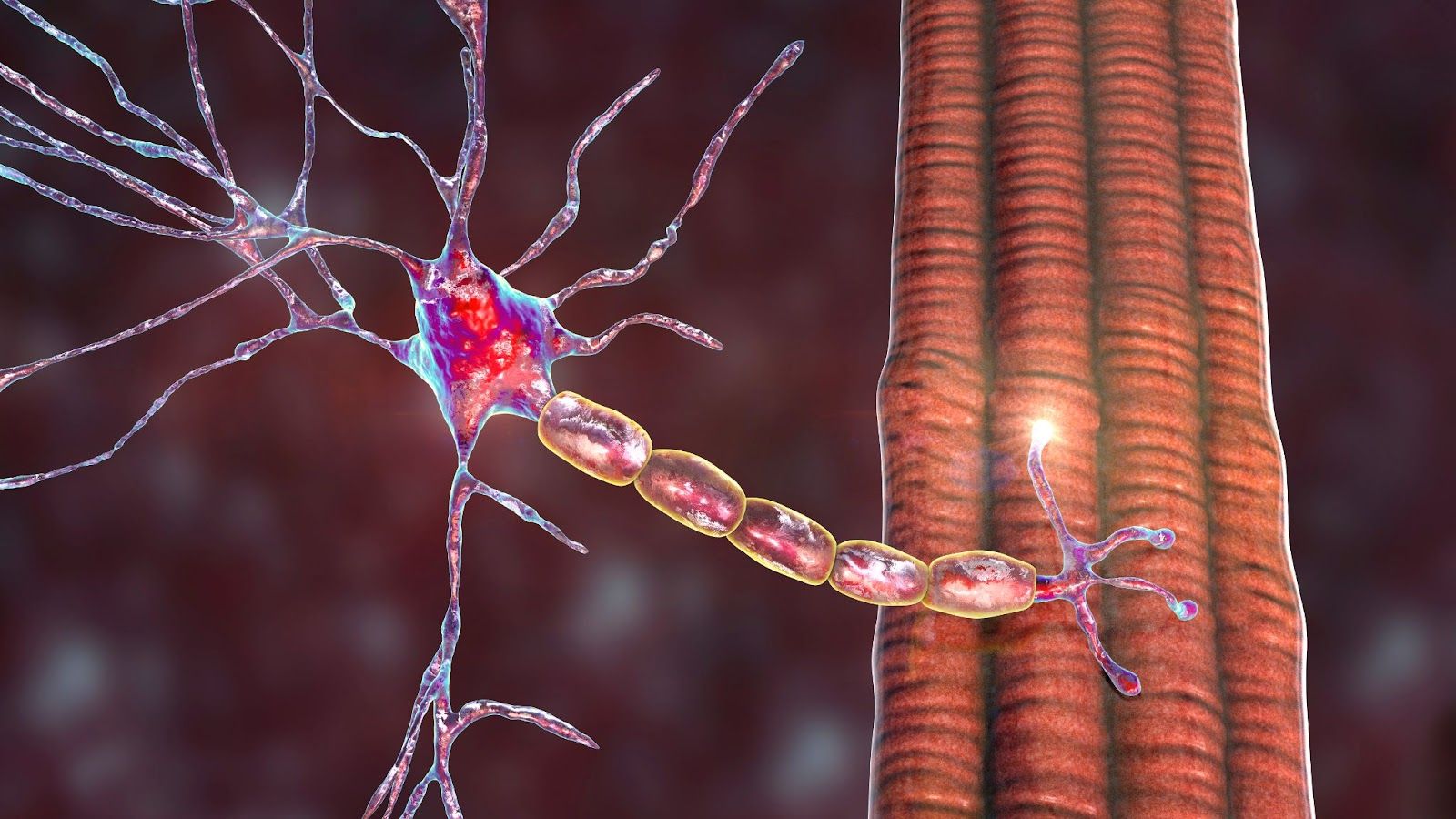
Image courtesy of Dr_Microbe
2. Detailed Structure of Neuromuscular Junctions
NMJs are composed of several distinct structures, each contributing to their unique function.
2.1 Presynaptic Terminal
Practice Questions
FAQ
The basal lamina at the neuromuscular junction (NMJ) plays multiple crucial roles in both structural integrity and functional regulation. Structurally, it acts as a scaffold, maintaining the alignment between the motor neuron terminal and the muscle fibre. This precise alignment is essential for effective neurotransmitter communication. Functionally, the basal lamina contains high concentrations of acetylcholinesterase (AChE), the enzyme responsible for breaking down acetylcholine (ACh) after it has been released into the synaptic cleft. This ensures rapid termination of the signal to prevent prolonged muscle contraction. Additionally, the basal lamina plays a role in the development and regeneration of the NMJ. It contains various signaling molecules and growth factors that contribute to the formation and maintenance of the junction, ensuring its functionality and plasticity. Thus, the basal lamina is a critical component, serving both as a structural anchor and a modulator of synaptic activity at the NMJ.
A neuromuscular junction (NMJ) is a unidirectional synapse, meaning it transmits signals only in one direction - from the motor neuron to the muscle fibre. This directionality is ensured by the specific arrangement and functioning of its components. The motor neuron's presynaptic terminal contains synaptic vesicles filled with acetylcholine (ACh), which are released into the synaptic cleft upon receiving an action potential. The muscle fibre's postsynaptic membrane has nicotinic ACh receptors that respond to ACh but does not release any neurotransmitters itself. Moreover, the NMJ lacks the mechanisms for reverse signal transmission, such as receptors on the motor neuron terminal or neurotransmitter release from the muscle fibre. This unidirectional nature is fundamental for the precise control of muscle contraction, as it ensures a clear and specific pathway for signal transmission, allowing for coordinated muscle movement.
If a neuromuscular junction (NMJ) is blocked or malfunctions, it can lead to serious consequences in muscle function, ranging from muscle weakness to complete paralysis. Blocking of NMJs can occur due to various factors, including toxins, autoimmune diseases, or certain medications. For instance, botulinum toxin (found in botulism) inhibits ACh release, preventing muscle contraction, which can lead to paralysis. In autoimmune disorders like myasthenia gravis, antibodies attack ACh receptors, reducing their number or function, leading to muscle weakness and fatigue. Certain anaesthetics and muscle relaxants used in surgery also work by temporarily blocking NMJs. When NMJs malfunction, the affected muscles cannot receive proper signals from the motor neurons, resulting in impaired muscle contractions. This can affect a range of activities, from simple movements to vital functions like breathing, depending on the muscles involved. Understanding and treating conditions affecting NMJs are therefore crucial in clinical neurology and anesthesiology.
Muscle relaxation at the neuromuscular junction (NMJ) is facilitated primarily by the cessation of acetylcholine (ACh) release and its rapid breakdown. When the motor neuron stops firing action potentials, it halts the release of ACh into the synaptic cleft. Concurrently, acetylcholinesterase (AChE), abundant in the synaptic cleft, degrades any remaining ACh. This enzymatic breakdown prevents further activation of nicotinic ACh receptors on the motor end plate, thus stopping the influx of sodium ions into the muscle fibre. The cessation of sodium ion influx halts further depolarisation of the muscle membrane. Subsequently, the muscle fibre repolarises, restoring the electrical gradient across the muscle cell membrane. This repolarisation is a critical step in muscle relaxation. Additionally, active transport mechanisms work to re-establish ion concentration gradients disrupted during contraction. Calcium ions are pumped back into the sarcoplasmic reticulum, and sodium-potassium pumps restore the original ion distribution, ensuring the muscle fibre returns to its resting state, ready for the next contraction.
Acetylcholinesterase (AChE) at the neuromuscular junction (NMJ) is crucial for the rapid and precise termination of muscle contraction signals. Located within the synaptic cleft, AChE swiftly breaks down the neurotransmitter acetylcholine (ACh) immediately after it binds to the nicotinic receptors on the motor end plate. This enzymatic breakdown is essential to prevent continuous stimulation of muscle fibres, which would lead to prolonged contraction and potential muscle fatigue. By efficiently hydrolyzing ACh into acetate and choline, AChE ensures that the neurotransmitter does not persist in the synaptic cleft and therefore avoids repetitive or sustained muscle contraction. This rapid clearing of ACh from the synaptic cleft allows the muscle fibre to return to its resting state, readying it for the next signal. The efficiency of AChE is so high that the ACh is typically broken down within milliseconds, exemplifying the highly coordinated nature of muscular control at the NMJ.
