
Introduction to Gel Electrophoresis
Gel electrophoresis is a fundamental technique in molecular biology, biochemistry, and genetics. This method separates DNA, RNA, or proteins on the basis of their size and charge. The technique is integral for DNA analysis, enabling scientists to study genetic material in detail.
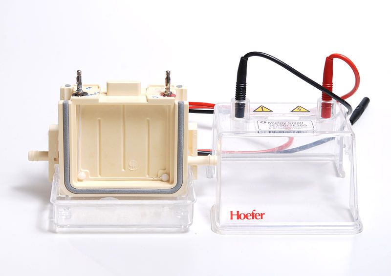
Polyacrylamide gel electrophoresis apparatus
Image courtesy of Lilly_M
Principles of Gel Electrophoresis
Gel electrophoresis is based on the movement of charged molecules in an electric field. DNA fragments are negatively charged due to their phosphate backbone and, thus, migrate towards the positive electrode in an electric field.
Components of Gel Electrophoresis
Practice Questions
FAQ
A molecular weight marker, also known as a DNA ladder, is essential in gel electrophoresis for determining the size of DNA fragments in the sample. These markers consist of DNA fragments of known lengths, providing a reference scale against which the sizes of unknown DNA fragments can be compared. By running the marker alongside the DNA samples, it becomes possible to estimate the size of the DNA fragments based on their relative mobility compared to the marker bands. This comparison is crucial for accurate analysis of gel electrophoresis results, especially in research and diagnostics where fragment size is a critical factor.
In gel electrophoresis, the electric field is fundamental to the migration of DNA fragments. DNA molecules are negatively charged due to their phosphate backbone, so when an electric field is applied, these molecules are attracted towards the positively charged electrode (anode). The strength of the electric field influences the rate of migration; a stronger field causes faster movement of DNA fragments. However, very high field strengths can cause overheating and may distort the gel structure, thus affecting the separation quality. Therefore, the electric field must be optimally maintained to ensure efficient and accurate separation of DNA fragments.
The buffer system in gel electrophoresis, such as TAE or TBE, plays a crucial role in maintaining the integrity of DNA samples. It provides a conducive environment by maintaining a stable pH, which is essential to prevent DNA degradation during electrophoresis. The buffer also facilitates the conduction of the electric current throughout the gel, ensuring a uniform electric field. This uniformity is important for consistent DNA migration. Furthermore, the ions in the buffer solution help to dissipate heat generated during electrophoresis, preventing damage to the DNA samples and the gel.
The loading dye in gel electrophoresis serves two primary functions. Firstly, it increases the density of the DNA sample, ensuring that the sample sinks into the wells of the gel and does not diffuse into the buffer. This is crucial for precise sample loading. Secondly, the dye provides colour, making the sample visible to the naked eye. This visibility allows for easier monitoring of the loading process and tracking the progress of DNA migration during electrophoresis. Additionally, the dye typically migrates at a predictable rate, acting as a rough indicator of how far the smallest DNA fragments have travelled.
Safety precautions during gel electrophoresis are crucial to prevent exposure to hazardous materials and electric shock. Ethidium bromide, commonly used for staining DNA, is a mutagen and should be handled with gloves and proper disposal methods. Additionally, since the process involves high-voltage electricity, care should be taken to avoid electric shock by turning off the power supply before handling the gel. UV light used for visualising DNA bands can damage eyes and skin, so protective eyewear and shields are necessary. Lastly, proper training and adherence to laboratory protocols are essential to ensure safe operation of the electrophoresis equipment.
