
24.2.2 X-ray Imaging
Mechanism of X-ray Imaging
X-ray imaging is a technique that uses X-rays to view the internal structure of an object, most commonly the human body. When X-rays are transmitted through the body, they are absorbed at varying degrees by different tissues due to their different densities. Bones, being denser, absorb more X-rays, making them appear white on the radiograph, while muscles and other soft tissues, which absorb less, appear in varying shades of grey.
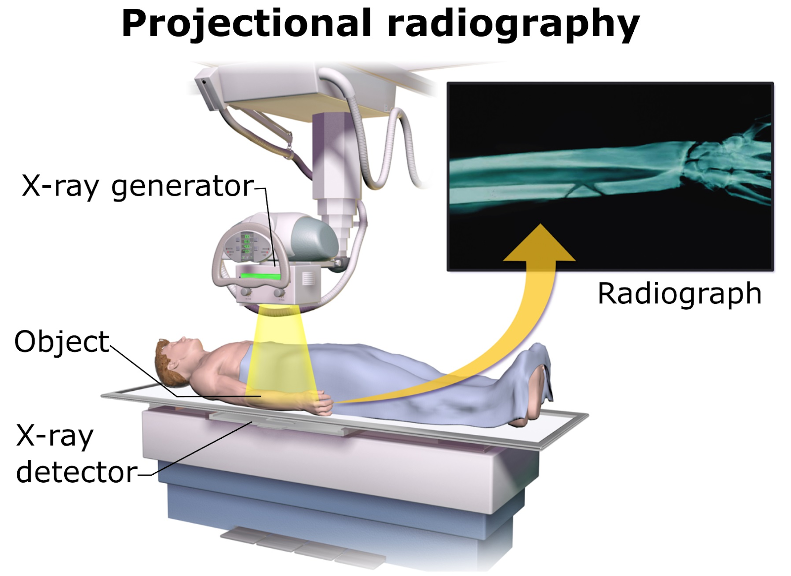
X-ray generator
Image Courtesy Blausen Medical
Key Components of X-ray Imaging Systems
Practice Questions
FAQ
The principle of X-ray attenuation is fundamental to the formation of an X-ray image. Attenuation refers to the reduction in intensity of the X-ray beam as it passes through different types of tissues in the body. Different tissues attenuate X-rays to varying degrees based on their density and composition. Dense tissues like bone have a high attenuation rate and absorb more X-rays, leading to fewer X-rays reaching the detector in these areas. This results in bones appearing white on an X-ray image. Conversely, less dense tissues like muscles or organs allow more X-rays to pass through, resulting in darker areas on the image. The varying degrees of attenuation across different tissues create the contrasts seen in an X-ray image, which are essential for diagnosing various conditions. Understanding how different tissues attenuate X-rays allows radiologists to interpret these images accurately.
Yes, X-ray imaging is not limited to detecting fractures and bone-related issues; it is also effective in diagnosing various other conditions. While X-rays are best known for their ability to visualize bone structures clearly, they can also be used to detect abnormalities in soft tissue and organs. For instance, X-rays are commonly used in diagnosing lung conditions such as pneumonia, where the infected areas appear as cloudy regions on the X-ray film. They are also used in dentistry to detect tooth decay and in mammography for breast cancer screening. However, the effectiveness of X-rays in diagnosing soft tissue conditions is somewhat limited compared to other imaging modalities like MRI or CT scans. These other techniques provide better contrast and detail for soft tissues, making them more suitable for certain diagnostic purposes. Nevertheless, X-rays remain a valuable and frequently used diagnostic tool in various medical fields due to their availability, speed, and cost-effectiveness.
While contrast media significantly enhance the diagnostic capabilities of X-ray imaging, there are several limitations to their use. One of the primary concerns is the risk of allergic reactions, which, although rare, can be severe. Patients with a history of allergies or kidney problems are at higher risk. To address this, pre-screening for allergies and renal function tests are often conducted. Another limitation is that contrast media can obscure certain structures, making it challenging to view adjacent tissues or organs. To mitigate this, precise dosing and timing of contrast administration are crucial. Additionally, the use of contrast media can be limited in patients with certain medical conditions, such as impaired kidney function, as it can exacerbate these conditions. In such cases, alternative imaging modalities or contrast agents with lower toxicity may be considered. Despite these limitations, contrast media remain an essential tool in enhancing the clarity and diagnostic value of X-ray images, with ongoing research aimed at developing safer and more effective contrast agents.
The disposal of X-ray films and associated chemicals used in traditional X-ray imaging poses significant environmental and safety concerns. X-ray films contain silver, a heavy metal, which can be harmful to the environment if not properly disposed of. The development and fixing solutions used in film processing contain chemicals that can be toxic and polluting. It's essential to follow strict guidelines for disposal to avoid environmental contamination. Specialised recycling programs exist for X-ray films where the silver is recovered and reused, reducing the environmental impact. The chemicals used in film processing must be treated as hazardous waste and disposed of accordingly. Many healthcare facilities now use digital X-ray systems, which eliminate the need for these hazardous materials, thereby significantly reducing the environmental footprint and enhancing safety in X-ray imaging.
Digital X-ray detectors offer significant advantages over traditional film-based detectors, primarily in image quality and radiation exposure. Digital detectors are more sensitive to X-rays, which means they can produce high-quality images at lower radiation doses. This reduction in exposure is crucial for patient safety, especially in cases where multiple scans are necessary. In terms of image quality, digital detectors provide sharper images with better contrast resolution. They also allow for immediate viewing and manipulation of images, such as adjusting brightness and contrast or zooming in for detail, which is not possible with film. Additionally, digital systems offer the advantage of storing and transferring images electronically, facilitating easier and quicker sharing among healthcare professionals. This capability enhances collaborative diagnosis and treatment planning. Overall, the transition to digital X-ray technology represents a significant leap in both diagnostic capabilities and patient safety in medical imaging.
