
Monomers, Polymers and Biological Reactions
The shared biochemical basis of life provides indirect evidence that living organisms have common evolutionary origins.
A monomer is a small molecular unit from which a larger molecule can be constructed.
A polymer contains many repeating monomers joined by chemical bonds.
Monosaccharides, amino acids and nucleotides are biologically important monomers.
A condensation reaction forms a chemical bond between molecules and eliminates one molecule of water.
A hydrolysis reaction uses one molecule of water to break a chemical bond.
Condensation constructs larger biological molecules, whereas hydrolysis separates them into smaller units.
Glucose Isomers and Glycosidic Bonds
Glucose occurs as the structural isomers α-glucose and β-glucose.
The isomers differ in the position of the hydroxyl group attached to carbon 1.
In α-glucose, the hydroxyl group on carbon 1 is below the plane of the ring.
In β-glucose, the hydroxyl group on carbon 1 is above the plane of the ring.
Condensation between glucose molecules forms glycosidic bonds.
The glucose isomer and glycosidic bonds present determine the structure and properties of the resulting polysaccharide.
Carbohydrate Tests and Analysis
For a reducing sugar, add Benedict’s solution and heat; a positive result changes from blue towards green, yellow, orange or brick-red.
For a non-reducing sugar, first boil with dilute acid, cool and neutralise before repeating Benedict’s test.
Add iodine in potassium iodide to test for starch; a positive result changes from orange-brown to blue-black.
In chromatography, compare separated sample spots with known monosaccharide standards to identify components.
A dilution series provides glucose solutions of known concentration.
React each standard under identical conditions and measure absorbance using a colorimeter.
Plot a calibration curve and use the unknown sample’s absorbance to determine its glucose concentration.
Phospholipids and the Emulsion Test
A phospholipid resembles a triglyceride, but one fatty acid is replaced by a phosphate-containing group.
Its phosphate-containing region is hydrophilic, whereas its fatty-acid tails are hydrophobic.
This contrast makes phospholipids suitable for forming the bilayer of plasma membranes.
Triglycerides are predominantly hydrophobic and function mainly as energy stores rather than membrane-forming molecules.
For the emulsion test, mix the sample with ethanol so any lipid dissolves.
Add the ethanol mixture to water; a cloudy white emulsion indicates lipid is present.
A negative result remains clear.
Levels of Protein Structure
Primary structure is the specific sequence of amino acids in a polypeptide.
Secondary structure is local folding into structures such as α-helices and β-pleated sheets, stabilised by hydrogen bonds.
Tertiary structure is the overall three-dimensional folding of one polypeptide.
Tertiary structure is stabilised by hydrogen bonds, ionic bonds and strong covalent disulfide bridges.
Quaternary structure is the association of two or more polypeptide chains into one functional protein.
The primary structure determines folding, and the final three-dimensional shape determines the protein’s properties and function.
Proteins function as cell structures, enzymes, chemical messengers and components of blood.
Enzyme Rate Factors and Practical Skills
Increasing enzyme concentration increases rate when sufficient substrate is available; increasing substrate concentration eventually reaches a maximum rate as active sites become saturated.
Increasing temperature raises collision frequency until the optimum; higher temperatures disrupt bonds, altering the active site and causing denaturation.
Extreme pH changes ionic and hydrogen bonding, altering tertiary structure and reducing activity.
A competitive inhibitor binds to the active site; increasing substrate concentration reduces its effect.
A non-competitive inhibitor binds elsewhere, altering the active site; additional substrate does not overcome its effect.
Calculate pH using pH = −log₁₀[H⁺].
In the required practical, vary one named factor, control other variables and repeat measurements to assess uncertainty.
Plot suitable graphs and calculate the initial rate from the gradient of a tangent at the start of the reaction.
Monosaccharides and Disaccharides
Monosaccharides are carbohydrate monomers; examples include glucose, galactose and fructose.
Two monosaccharides join by condensation, forming a glycosidic bond and releasing water.
A disaccharide consists of two monosaccharides joined by a glycosidic bond.
Maltose forms from two glucose molecules.
Sucrose forms from glucose and fructose.
Lactose forms from glucose and galactose.
Hydrolysis reverses disaccharide formation by breaking the glycosidic bond using water.
Starch, Glycogen and Cellulose
Polysaccharides form through repeated condensation reactions between many glucose molecules.
Starch is an α-glucose storage polymer in plants, consisting of amylose and amylopectin.
Amylose is unbranched and coils compactly; amylopectin is branched, providing many ends for rapid hydrolysis.
Glycogen is a compact, highly branched α-glucose polymer used for carbohydrate storage in animal cells.
Its extensive branching provides many ends where enzymes can release glucose rapidly.
Cellulose contains straight, unbranched chains of β-glucose with alternate molecules inverted.
Hydrogen bonds between cellulose chains form strong microfibrils, giving plant cell walls tensile strength.
Triglycerides and Fatty Acids
A triglyceride forms from one glycerol molecule and three fatty acids through three condensation reactions.
Each condensation forms an ester bond between glycerol and a fatty acid and releases water.
A saturated fatty acid has no carbon–carbon double bonds.
An unsaturated fatty acid contains at least one carbon–carbon double bond, which introduces a bend into the chain.
Triglycerides are non-polar and hydrophobic, so they do not affect the water potential of cells.
Their high ratio of carbon–hydrogen bonds makes them concentrated respiratory energy stores.
Their insolubility allows compact storage without loss by diffusion from cells.
Amino Acids, Peptide Bonds and Protein Tests
An amino acid contains an amine group, a carboxyl group, a hydrogen atom and an R group attached to a central carbon.
The 20 amino acids common to organisms differ only in their R groups.
Condensation between two amino acids forms a peptide bond and releases water.
Two joined amino acids form a dipeptide; many joined amino acids form a polypeptide.
A functional protein can contain one polypeptide or several associated polypeptides.
For the Biuret test, add Biuret reagent; a lilac or purple colour indicates peptide bonds.
Chromatography with known standards can separate and identify amino acids in a mixture.
Enzyme Action and Specificity
An enzyme increases reaction rate by lowering the reaction’s activation energy.
The enzyme’s active site is determined by its tertiary structure and can bind complementary substrates.
In the induced-fit model, substrate binding causes the active site to change shape, improving its fit around the substrate.
An enzyme–substrate complex forms, bonds are altered and products are released.
The active site then returns to a form capable of catalysing another reaction.
Enzyme specificity results from the active site’s complementary shape and chemical properties.
Enzyme-action models have changed as new evidence showed that active sites are flexible rather than completely rigid.
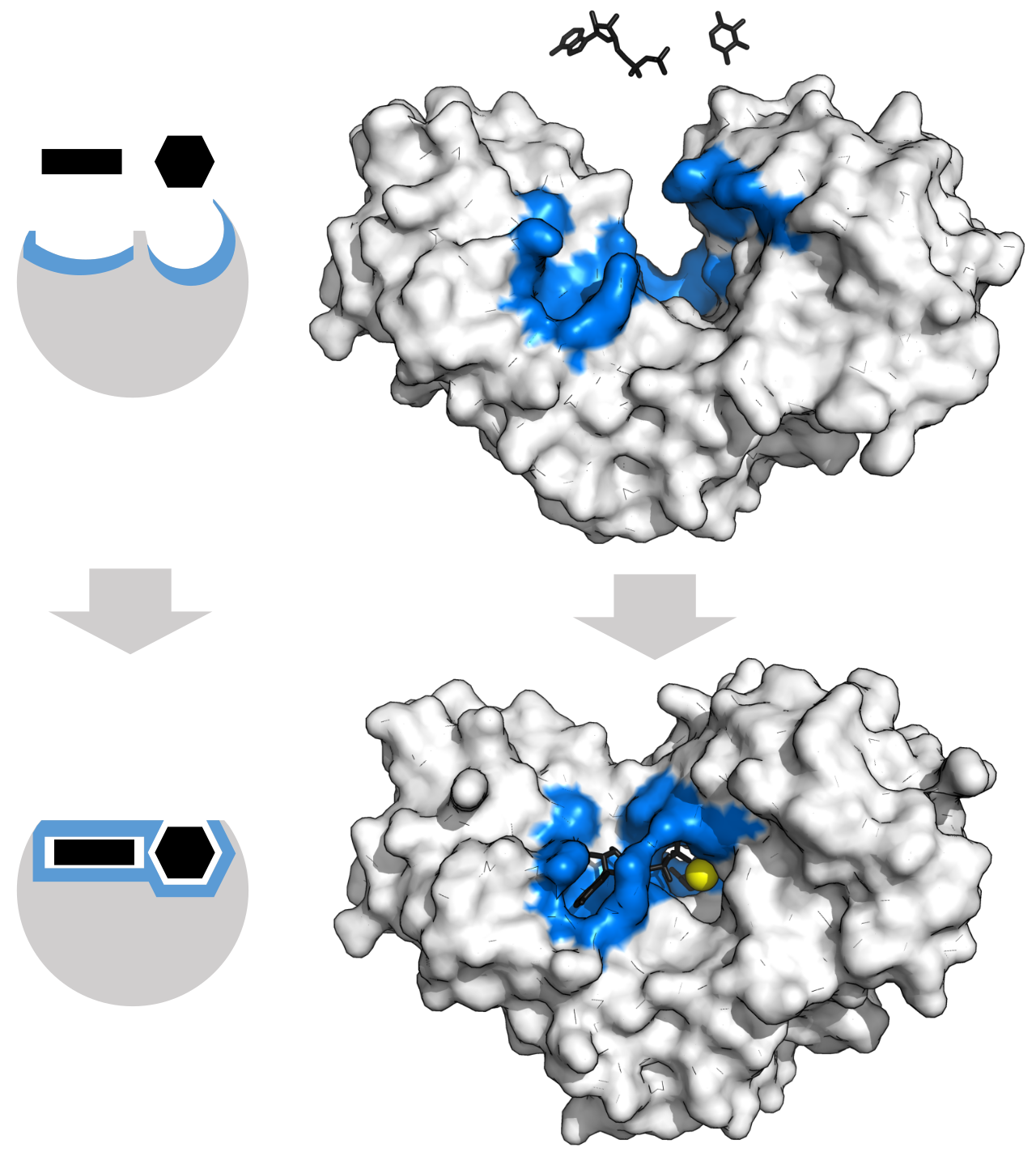
Substrate binding causes the enzyme’s active site to change conformation and close around the substrate. This illustrates why active sites are flexible and how an enzyme–substrate complex forms. Source
Checklist: can you do this?
Can you distinguish condensation from hydrolysis and identify the bond formed in each biological molecule?
Can you explain how the structures of starch, glycogen, cellulose, triglycerides and phospholipids suit their functions?
Can you interpret the results of Benedict’s, iodine, emulsion and Biuret tests?
Can you predict how substrate concentration, temperature, pH and inhibitors affect enzyme-controlled reaction rates?
Can you calculate pH, use a calibration curve and determine an initial rate from a tangent?