The circulatory system
· Mammalian circulatory system = closed double circulation.
· Closed circulation = blood remains inside blood vessels.
· Double circulation = blood passes through the heart twice during one complete circuit: pulmonary circulation + systemic circulation.
· Main parts: heart, blood, blood vessels.
· Blood vessels include arteries, arterioles, capillaries, venules and veins.
· Advantage of double circulation: separates oxygenated and deoxygenated blood and maintains high pressure to body tissues.
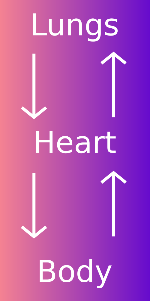
This diagram shows how mammals have two linked circuits: one between the heart and lungs, and one between the heart and body tissues. It is useful for identifying the idea of closed double circulation and the direction of blood flow. Source
Pulmonary and systemic circulation
· Pulmonary circulation = blood flow between heart and lungs.
· Pulmonary artery carries deoxygenated blood from the right ventricle to the lungs.
· Pulmonary vein carries oxygenated blood from the lungs to the left atrium.
· Systemic circulation = blood flow between heart and body tissues.
· Aorta carries oxygenated blood from the left ventricle to the body.
· Vena cava carries deoxygenated blood from the body to the right atrium.
· Exam tip: arteries carry blood away from the heart; veins carry blood towards the heart — this is true even if the blood is deoxygenated, e.g. pulmonary artery.
Blood vessel structure and function
· Arteries carry blood away from the heart at high pressure.
· Muscular arteries have a thick tunica media with much smooth muscle to control blood flow by vasoconstriction and vasodilation.
· Elastic arteries have many elastic fibres to stretch during systole and recoil during diastole, smoothing blood flow and maintaining pressure.
· Arterioles are small arteries that regulate blood flow into capillary beds by changing lumen diameter.
· Capillaries are the site of exchange between blood and tissues.
· Capillary walls are one cell thick, made of endothelium, giving a short diffusion distance.
· Capillaries have a very narrow lumen so red blood cells pass slowly and close to the wall, increasing time for diffusion.
· Venules collect blood from capillaries and join to form veins.
· Veins carry blood towards the heart at low pressure.
· Veins have a large lumen, thin wall, less smooth muscle and fewer elastic fibres than arteries.
· Veins contain valves to prevent backflow of blood.
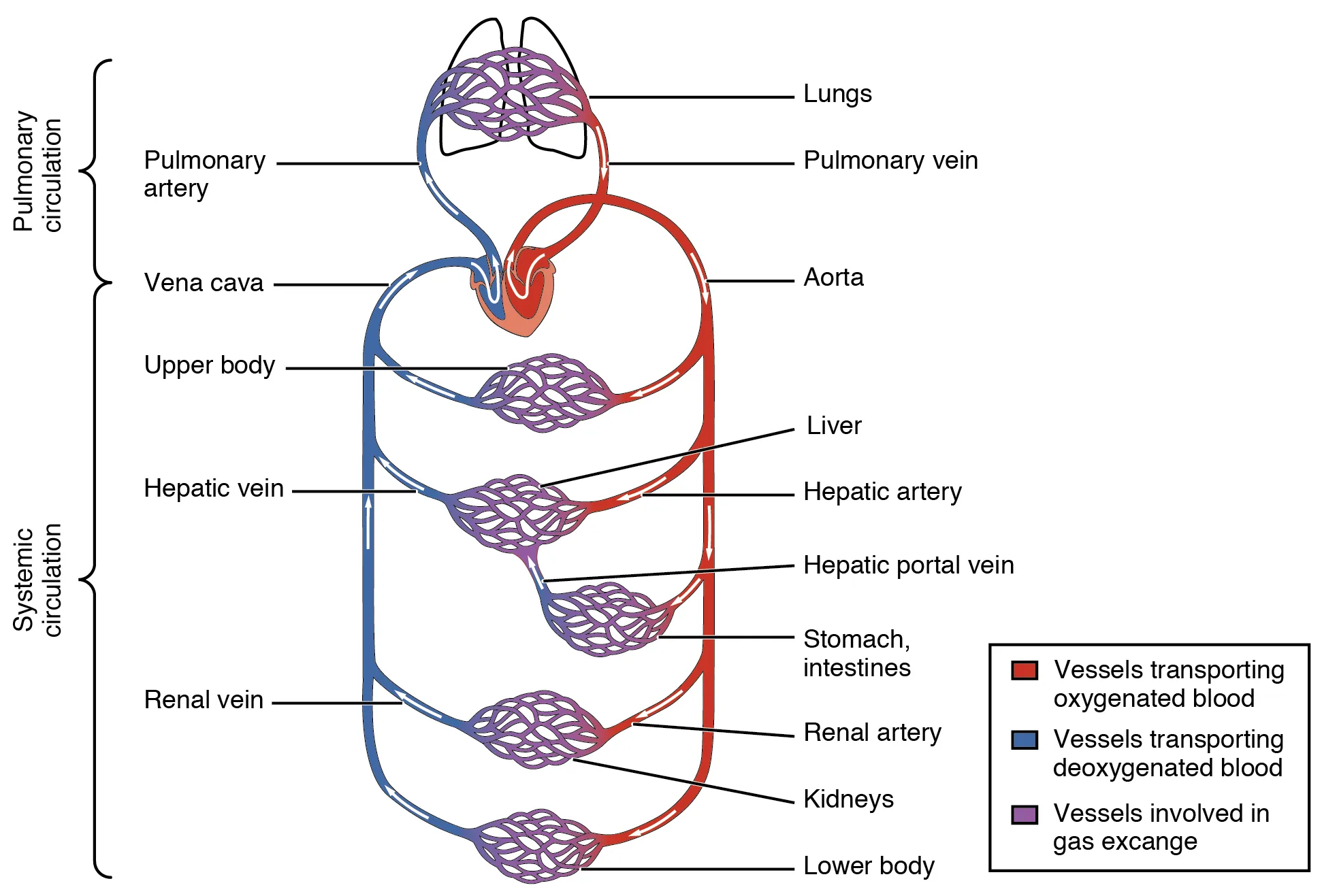
This diagram compares the structure of arteries, veins and capillaries. It is useful for linking wall thickness, elastic tissue, smooth muscle, lumen size and valves to each vessel’s function. Source
Recognising arteries, veins and capillaries in images
· Artery TS: thick wall, small round lumen, prominent smooth muscle and elastic fibres.
· Vein TS: thinner wall, larger irregular lumen, may contain valves.
· Capillary: very small vessel, wall one endothelial cell thick, often just wide enough for one red blood cell.
· In longitudinal section, arteries and veins appear as tubes; identify by wall thickness, lumen size and presence or absence of valves.
· Plan diagrams should show outline only, no shading, and should label major tissues clearly.
· For arteries and veins in TS, label lumen, tunica intima/endothelium, tunica media, tunica externa where visible.
· Exam skill: always relate visible structure to function, e.g. thick elastic wall withstands high pressure.
Blood cells to recognise and draw
· Red blood cells / erythrocytes: small biconcave discs, no nucleus, pale centre in microscope images; transport oxygen using haemoglobin.
· Neutrophils: white blood cells with a multi-lobed nucleus and granular cytoplasm; involved in phagocytosis.
· Monocytes: large white blood cells with a kidney-shaped nucleus; can develop into macrophages.
· Lymphocytes: white blood cells with a large round nucleus and thin rim of cytoplasm; important in specific immunity.
· In drawings, use clear outlines and label distinctive features such as nucleus shape, cell size and presence/absence of granules.
· Do not confuse red blood cells with lymphocytes: red blood cells are smaller, biconcave and lack a nucleus.
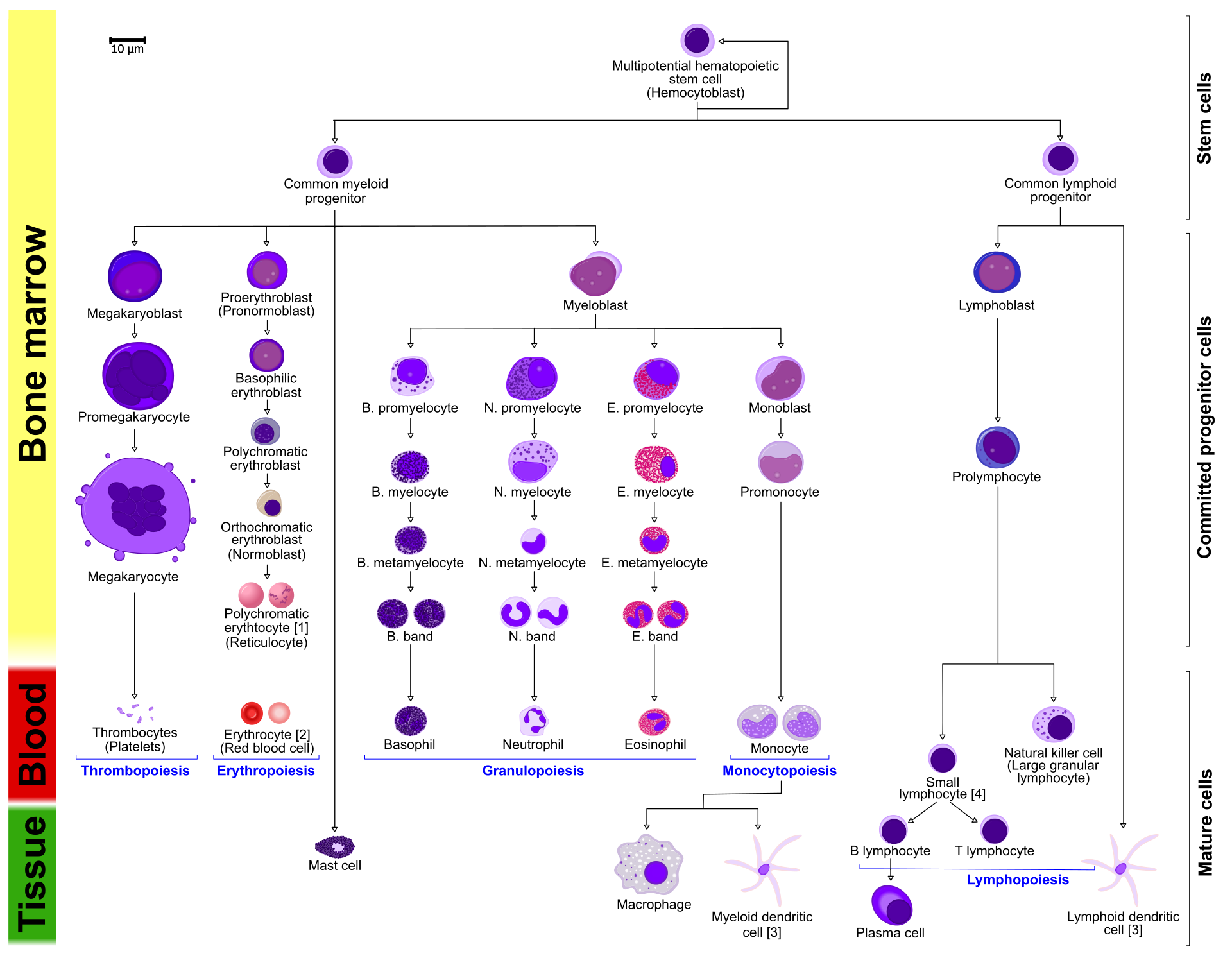
This diagram helps compare the appearance of major blood cell types. Focus on the features needed for recognition in microscope images: red blood cells lack nuclei, while white blood cells differ in nucleus shape and cytoplasm. Source
Water, blood and tissue fluid
· Water is the main component of blood and tissue fluid.
· Solvent action: water dissolves many substances, allowing transport of glucose, amino acids, ions, hormones, carbon dioxide and urea in blood plasma.
· High specific heat capacity: water resists temperature change, helping blood distribute heat and maintain a stable internal temperature.
· Tissue fluid surrounds body cells and allows exchange between blood and cells.
· Tissue fluid supplies cells with oxygen, glucose, amino acids, ions and other useful substances.
· Tissue fluid removes carbon dioxide and other metabolic wastes from cells.
Formation of tissue fluid
· Tissue fluid forms in capillary networks by filtration from blood plasma.
· At the arterial end of a capillary, hydrostatic pressure is high because blood enters from arterioles.
· High hydrostatic pressure forces plasma out through gaps in the capillary wall, forming tissue fluid.
· Large proteins and blood cells usually remain in the capillary because they are too large to pass through the capillary wall.
· Tissue fluid bathes cells, allowing diffusion of useful substances into cells and wastes out of cells.
· At the venous end, hydrostatic pressure is lower, so some tissue fluid returns to the capillary.
· Excess tissue fluid drains into the lymphatic system and eventually returns to the blood.
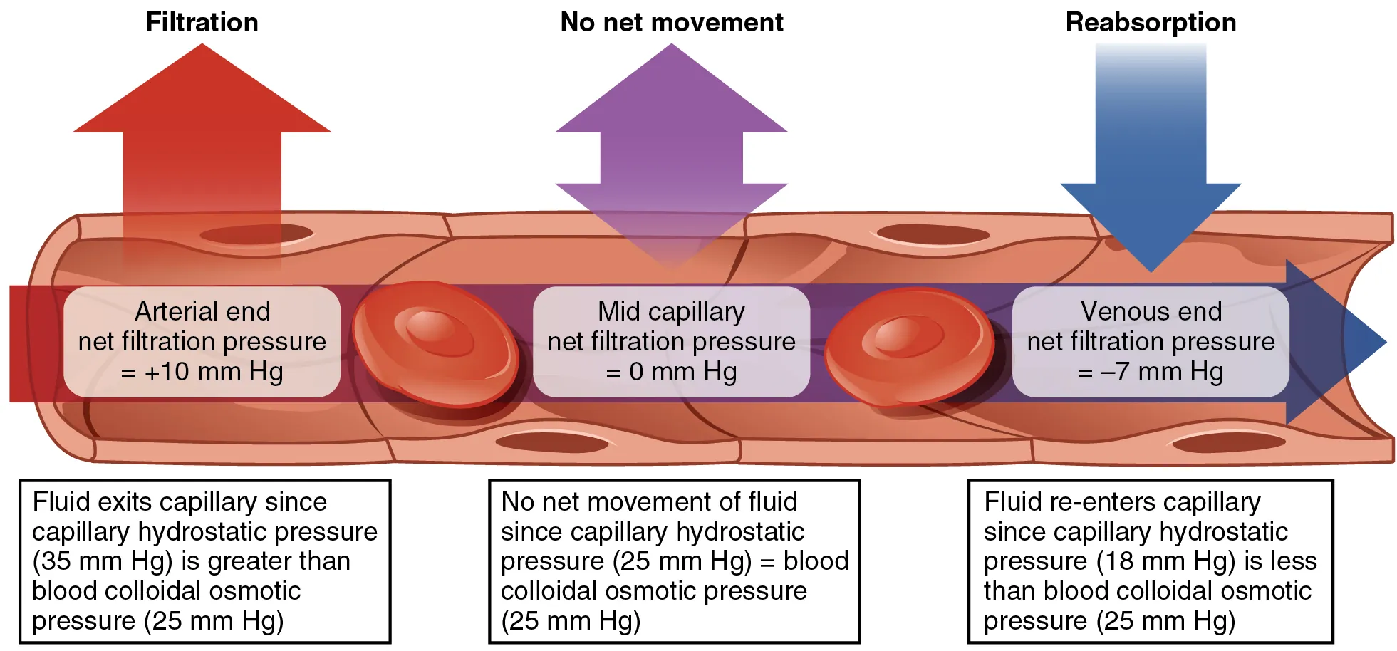
This diagram shows how hydrostatic pressure and osmotic effects control movement of fluid across capillary walls. It supports exam answers on how tissue fluid forms and why some fluid returns to the blood at the venous end. Source
Structure-function links to learn
· Artery thick wall → withstands high pressure from the heart.
· Elastic fibres in arteries → stretch and recoil to maintain continuous blood flow.
· Smooth muscle in muscular arteries/arterioles → changes lumen diameter to control blood distribution.
· Vein large lumen → reduces resistance to blood flow at low pressure.
· Vein valves → prevent backflow.
· Capillary one-cell-thick wall → short diffusion distance for exchange.
· Capillary narrow lumen → slows blood flow and keeps red blood cells close to exchange surface.
· Many capillaries → large surface area for exchange.
Common exam mistakes
· Do not say the pulmonary artery carries oxygenated blood; it carries deoxygenated blood to the lungs.
· Do not say all arteries carry oxygenated blood; arteries carry blood away from the heart.
· Do not say capillaries have thick walls; capillaries have walls one endothelial cell thick.
· Do not confuse tissue fluid with blood plasma; tissue fluid lacks red blood cells and has very little protein.
· Do not describe vessel structure without linking it to function.
· Do not over-detail heart structure or oxygen transport here; those are mainly 8.2 and 8.3, not 8.1.
Checklist: can you do this?
· State that mammals have a closed double circulation made of heart, blood and blood vessels.
· Describe the functions of the pulmonary artery, pulmonary vein, aorta and vena cava.
· Recognise and draw arteries, veins, capillaries, red blood cells, monocytes, neutrophils and lymphocytes from images.
· Explain how vessel structures are adapted to function, especially arteries, elastic arteries, muscular arteries, veins and capillaries.
· Describe how tissue fluid forms in a capillary network and explain its function in exchange.