External structure of the mammalian heart
· The mammalian heart is a muscular organ that pumps blood through pulmonary circulation and systemic circulation.
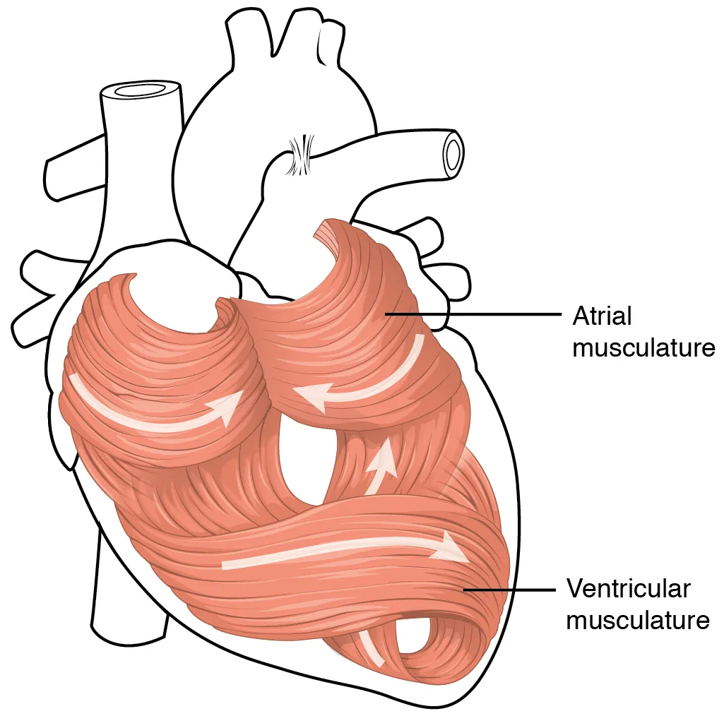
· The heart is surrounded by cardiac muscle, which contracts rhythmically and does not fatigue easily.
· The main external blood vessels are: aorta, vena cava, pulmonary artery and pulmonary vein.
· Vena cava carries deoxygenated blood from the body to the right atrium.
· Pulmonary artery carries deoxygenated blood from the right ventricle to the lungs.
· Pulmonary vein carries oxygenated blood from the lungs to the left atrium.
· Aorta carries oxygenated blood from the left ventricle to the body.
· In diagrams, remember: the right side of the heart is usually shown on the left side of the page.
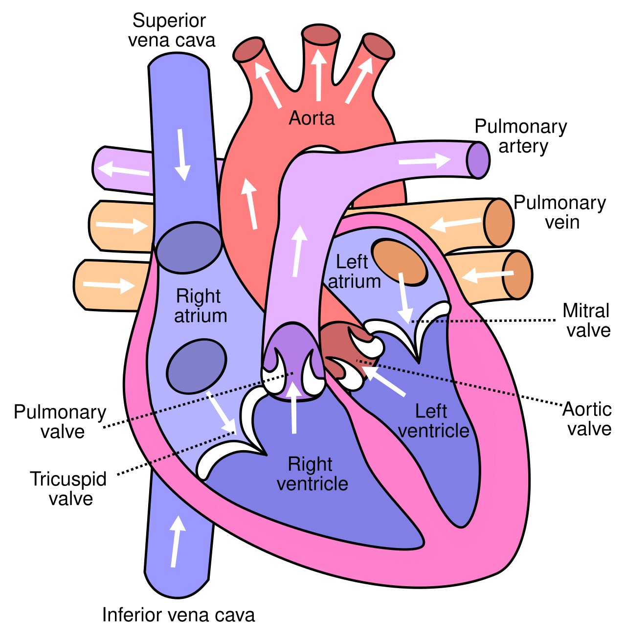
This diagram shows the main structures of the human heart, including the atria, ventricles, valves and great vessels. It is useful for learning blood flow routes and identifying key structures in exam diagrams. Source
Internal structure of the heart
· The heart has four chambers: right atrium, right ventricle, left atrium, left ventricle.
· Atria receive blood entering the heart; ventricles pump blood out of the heart.
· The septum separates the left and right sides of the heart, preventing mixing of oxygenated and deoxygenated blood.
· Atrioventricular valves are between atria and ventricles: tricuspid valve on the right, bicuspid / mitral valve on the left.
· Semilunar valves are found at the bases of the aorta and pulmonary artery.
· Valves ensure one-way flow of blood by opening and closing due to pressure differences.
· Tendinous cords prevent the atrioventricular valves from turning inside out during ventricular contraction.
These diagrams show the internal anatomy of the heart and the major vessels entering and leaving it. They are useful for connecting chamber structure with direction of blood flow. Source
Thickness of heart walls
· Atria have thin muscular walls because they only pump blood a short distance into the ventricles.
· Ventricles have thicker muscular walls because they generate higher pressure to pump blood out of the heart.
· The left ventricle has the thickest wall because it pumps blood around the whole body through the systemic circulation.
· The right ventricle is thinner than the left because it only pumps blood to the lungs through the pulmonary circulation.
· The pressure from the right ventricle must be lower to avoid damaging delicate lung capillaries.
Cardiac cycle: key stages
· The cardiac cycle is one complete heartbeat, involving repeated contraction and relaxation of atria and ventricles.
· Systole = contraction; diastole = relaxation.
· During atrial systole, the atria contract and blood is pushed into the ventricles; atrioventricular valves are open.
· During ventricular systole, the ventricles contract; pressure rises, atrioventricular valves close, then semilunar valves open so blood leaves via the aorta and pulmonary artery.
· During diastole, the heart relaxes; pressure in ventricles falls, semilunar valves close, and blood flows into the atria and then into the ventricles.
· Blood always moves from higher pressure to lower pressure.
· Valve movements are caused by pressure changes, not by the valves actively contracting.
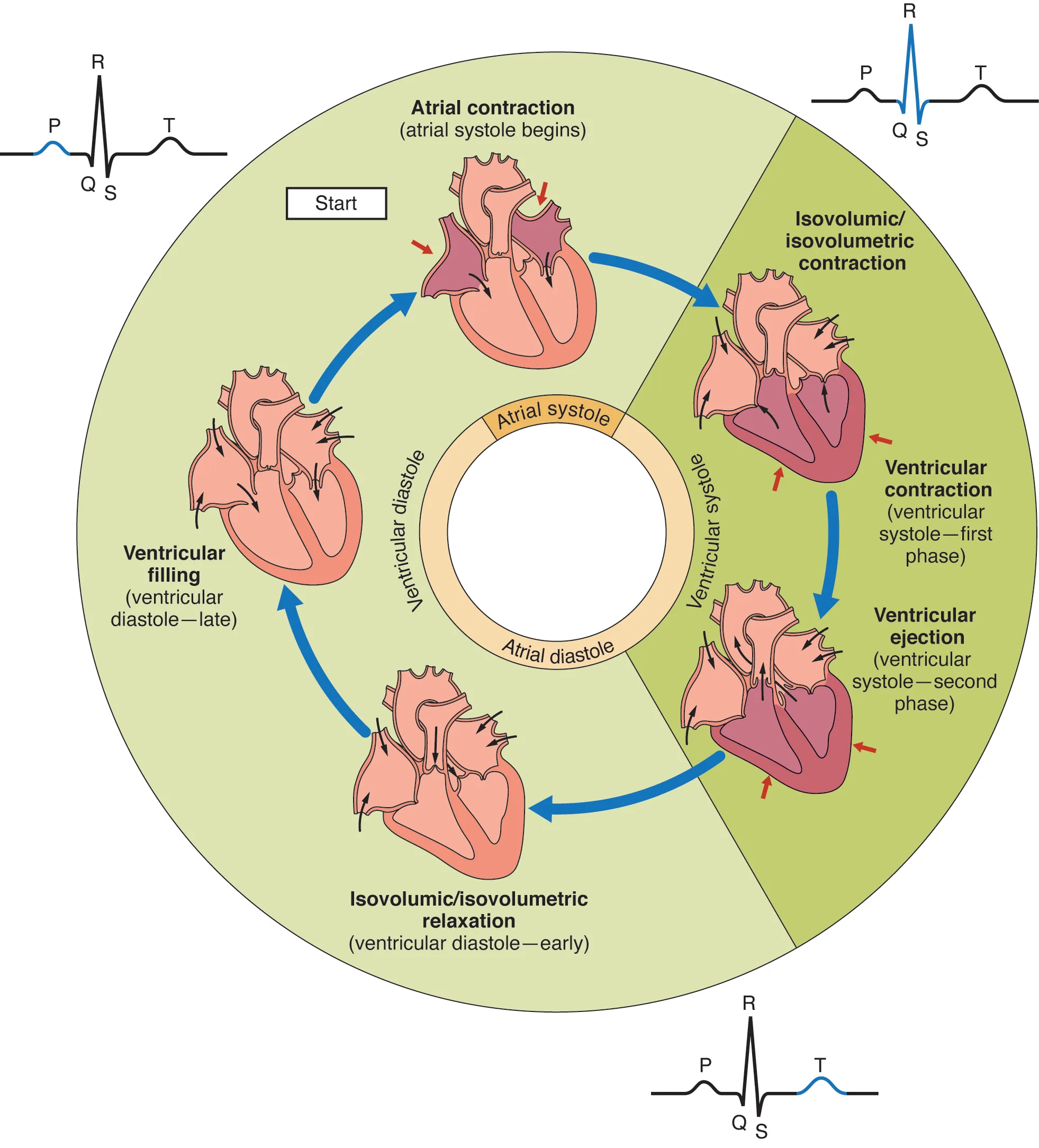
This diagram summarises how atrial systole, ventricular systole and diastole occur in sequence. It helps explain how pressure changes cause valve opening and closing during one heartbeat. Source
Pressure changes and valve action
· When atrial pressure > ventricular pressure, atrioventricular valves open and blood enters ventricles.
· When ventricular pressure > atrial pressure, atrioventricular valves close, producing the first heart sound.
· When ventricular pressure > pressure in arteries, semilunar valves open and blood is ejected.
· When arterial pressure > ventricular pressure, semilunar valves close, preventing backflow into ventricles.
· Valve closure prevents backflow and maintains efficient one-way circulation.
· Exam answers should link each valve movement to a specific pressure difference.
Electrical control of the cardiac cycle
· The heart is myogenic, meaning cardiac muscle initiates its own contraction without needing nervous stimulation.
· The sinoatrial node (SAN / SA node) acts as the pacemaker and initiates the wave of electrical excitation.
· The wave of excitation spreads across the atria, causing atrial systole.
· The atrioventricular node (AVN / AV node) delays the impulse briefly so the atria finish contracting before the ventricles contract.
· The impulse then travels down the Purkyne tissue / Purkinje fibres through the septum and into the ventricle walls.
· Purkyne tissue causes the ventricles to contract from the base upwards, pushing blood towards the aorta and pulmonary artery.
· For CIE 8.3, nervous and hormonal control are not required.
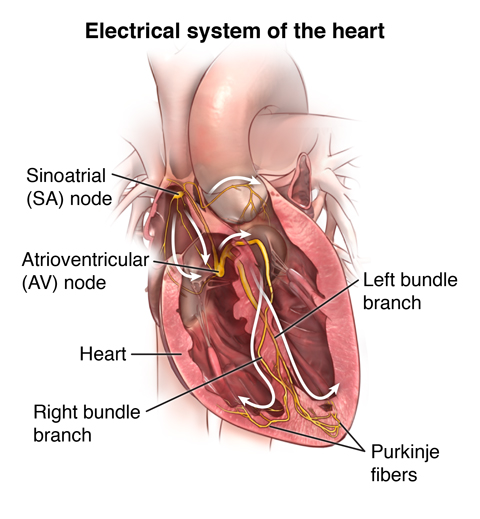
This diagram shows how electrical impulses spread through the heart to coordinate contraction. It is especially useful for understanding the roles of the SA node, AV node and Purkinje fibres in the cardiac cycle. Source
Common exam phrases
· “Left ventricle has thicker muscle because it pumps blood at high pressure around the body.”
· “Right ventricle has thinner muscle because it pumps blood only to the lungs.”
· “Valves open and close due to pressure differences.”
· “Atrioventricular valves prevent backflow from ventricles to atria.”
· “Semilunar valves prevent backflow from arteries to ventricles.”
· “SAN initiates excitation; AVN delays excitation; Purkyne tissue spreads excitation through ventricles.”
· “Ventricles contract from the base upwards to force blood into arteries.”
Checklist: can you do this?
· Label the main external and internal structures of the mammalian heart.
· Explain why atria, right ventricle and left ventricle have different wall thicknesses.
· Describe the cardiac cycle using systole, diastole, pressure changes and valve action.
· Explain the roles of the SAN, AVN and Purkyne tissue in coordinating heartbeat.
· Interpret diagrams showing chambers, valves, blood vessels, pressure changes or the conduction pathway.