
OCR Specification focus:
‘Examine stained sections or squashes and produce labelled diagrams of mitosis stages.’
Microscopic observation of mitosis in plant tissues allows biologists to visualise cell division, identify its distinct stages, and understand how genetic continuity is maintained across generations of cells.
Observing Mitosis in Plant Tissues
Studying mitosis in plants offers insight into how eukaryotic cells divide to form genetically identical daughter cells. This practical skill combines microscopy, slide preparation, and biological drawing to demonstrate understanding of the mitotic process. Plant root tips, particularly from onions (Allium cepa), are ideal because they contain actively dividing meristematic cells at their growing regions.
Choice of Material: Onion Root Tips
The root apical meristem is a zone of intense cell division, making it ideal for observing mitosis.
Cells here are small, with large nuclei, and divisions occur in quick succession. The uniform structure of onion roots simplifies observation and reduces variation between samples.
Preparation of Root Tip Squash Slides
A root tip squash enables clear viewing of chromosomes by flattening the tissue into a single cell layer under a coverslip.
Procedure Overview:
Fixation:
Root tips are cut and fixed (commonly in ethanoic alcohol or Carnoy’s fluid) to preserve cell structure and stop mitosis at various stages.Hydrolysis:
Root tips are softened by heating in hydrochloric acid (usually 1 mol dm⁻³ for several minutes). This breaks down the middle lamella, allowing cells to separate more easily.Staining:
The softened tissue is stained to highlight chromosomes.
Common stains include:Acetic orcein
Toluidine blue
Feulgen stain (binds specifically to DNA)
Squashing:
The stained root tip is placed on a slide, a coverslip added, and gently pressed (using folded paper to prevent slipping) to spread the cells into a thin layer for observation.
The goal is to produce a clear, evenly spread preparation where individual cells and chromosomes are visible.
Use of the Microscope
Once the slide is prepared:
Begin observation under low power to locate the meristematic region just behind the root cap.
Switch to high power (400× total magnification) to observe individual cells undergoing mitosis.
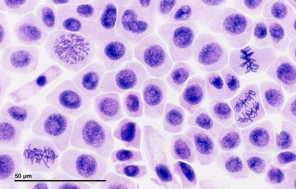
Onion root tip squash under bright-field microscopy showing cells in prophase, metaphase, anaphase, and telophase. The meristem provides many actively dividing cells, making stages easy to locate. This micrograph exemplifies what students should expect to see when scanning their own slides. Source.
Oil immersion objectives may be used for finer chromosomal detail in some laboratory settings.
Lighting and focus adjustments are critical to distinguishing fine structures like chromatid separation.
Stages of Mitosis in Plant Cells
Observation under the microscope allows identification of five key phases of mitosis, though cytokinesis may overlap with the later stages.
Interphase
Cells appear normal with an intact nuclear envelope and diffuse chromatin. The cell prepares for division by replicating DNA and organelles.
Interphase: The period between mitotic divisions when the cell grows, replicates DNA, and prepares for mitosis.
Chromosomes are not yet visible, but the nucleolus may be clearly seen.
Prophase
Chromatin condenses into visible chromosomes, each consisting of two sister chromatids joined at a centromere.
The nuclear envelope begins to break down.
Spindle fibres start to form from microtubules at opposite poles of the cell.
Metaphase
Chromosomes align at the cell equator, attached to spindle fibres via their centromeres.
This alignment ensures accurate chromosome segregation.
Metaphase: The stage of mitosis in which chromosomes line up along the equatorial plane of the cell.
Anaphase
Centromeres split, allowing sister chromatids to separate and move toward opposite poles.
The chromatids are pulled by the shortening of spindle fibres, creating characteristic V-shaped structures as they migrate.
Telophase
Chromatids (now individual chromosomes) reach opposite poles.
Nuclear envelopes reform around each set of chromosomes.
Chromosomes decondense, returning to thread-like chromatin.
Cytokinesis
In plants, a cell plate forms along the equator.
Vesicles from the Golgi apparatus fuse to create the new cell wall separating the two daughter cells.
Producing Labelled Diagrams
Students must produce accurate, clear biological drawings of each stage.
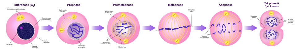
Schematic of prophase, metaphase, anaphase, and telophase/cytokinesis highlighting chromosome condensation, alignment, separation, and reformation of nuclei. This aids diagram quality and labelling expected in OCR drawings. Extra detail: the cleavage furrow shown is animal-cell-like; plant cells instead form a cell plate. Source.
These should:
Use sharp pencil lines (not shading or colouring).
Include labels for key structures: chromosomes, spindle fibres, nuclear envelope, and cell plate.
Maintain correct proportions and relative sizes.
Display one representative cell per stage, avoiding artistic embellishment.
Biological drawing: A precise, labelled scientific representation of observed biological structures, emphasising accuracy over artistry.
Drawings serve as a record of observation and demonstrate recognition of mitotic stages under the microscope.
Estimating the Mitotic Index
Quantifying mitosis is an important analytical step in this practical.
EQUATION
—-----------------------------------------------------------------
Mitotic Index (MI) = (Number of cells in mitosis ÷ Total number of cells observed) × 100
MI = percentage of dividing cells, indicating growth activity.
—-----------------------------------------------------------------
A high mitotic index signifies rapid cell division, typical of active growth regions, whereas a low value suggests slower division.
After calculating the mitotic index, students should consider sampling accuracy by counting cells across several fields of view to ensure reliability.
Practical Skills and Considerations
Safety:
Hydrochloric acid and stains can be corrosive; use gloves and goggles.Quality control:
Avoid over-squashing (which ruptures cells) or under-squashing (which prevents separation).Temperature control:
Excessive heating during hydrolysis damages tissue integrity.Data handling:
Record counts and mitotic indices neatly in tables, identifying each mitotic stage for clarity.
Good practice also includes repeat observations to verify consistency between slides.
Significance of Observing Mitosis
Observing mitosis in plant tissues provides essential understanding of cell cycle regulation, genetic stability, and the basis for growth and repair.
It reinforces theoretical learning by linking the visible stages of division to underlying molecular events such as DNA replication, chromosome alignment, and spindle function.
Practice Questions
Question 1 (2 marks)
When preparing a root tip squash to observe mitosis in onion cells, explain why the tissue is first treated with hydrochloric acid and then stained with a DNA-specific dye such as Feulgen stain.
Mark scheme:
1 mark for stating that hydrochloric acid softens the tissue or breaks down the middle lamella, allowing cells to separate easily.
1 mark for stating that the stain binds to DNA, making chromosomes visible under the microscope.
Question 2 (5 marks)
Describe how you would prepare and use a root tip squash to observe cells undergoing mitosis, and explain how you could calculate the mitotic index from your observations.
Mark scheme:
1 mark for cutting and fixing the root tip to preserve cells and halt mitosis at different stages.
1 mark for softening the tissue with hydrochloric acid to separate cells.
1 mark for staining the root tip with a DNA-specific dye (e.g. acetic orcein or toluidine blue) to make chromosomes visible.
1 mark for squashing the tissue gently under a coverslip to create a thin layer suitable for light microscopy.
1 mark for describing the mitotic index formula or method: counting the number of cells in mitosis divided by the total number of cells, multiplied by 100, to indicate growth activity.
FAQ
Onion root tips contain meristematic cells, which are regions of continuous growth where mitosis occurs frequently. These cells are small, actively dividing, and easy to stain, allowing all mitotic stages to be seen clearly.
Additionally, onion cells are large with distinct nuclei and chromosomes, and their lack of chloroplasts makes microscopic observation simpler. Root tips are also inexpensive and easy to handle, making them ideal for teaching and examination purposes.
Overheating during acid hydrolysis or staining can denature cellular proteins and damage chromosomal structure, making chromosomes appear blurred or fragmented.
This also causes cytoplasmic shrinkage or bursting of cells, which prevents accurate identification of mitotic stages.
To prevent this, heating should be gentle (around 60°C for a few minutes), ensuring the tissue softens without cellular distortion.
In late anaphase, chromatids are still moving apart but are visibly separated and migrating toward opposite poles. Spindle fibres are clearly visible, and the cell is elongated.
In early telophase, chromatids have reached the poles, and nuclear envelopes begin reforming around each set of chromosomes. Chromosomes also start to decondense, becoming less distinct.
A faint cell plate may begin to form in plant cells, signalling the transition to cytokinesis.
The squash technique flattens cells into a single layer, enabling clearer viewing of chromosomes and individual mitotic stages without complex equipment.
Benefits include:
Quicker and simpler than microtome sectioning.
Provides a larger field of view for counting cells.
Preserves mitotic detail when done gently.
However, over-squashing can rupture cells, so pressure must be applied carefully and evenly.
To improve reliability:
Count at least several hundred cells across multiple microscope fields.
Use random sampling to avoid selecting regions with abnormal division rates.
Clearly define what counts as a mitotic cell (visible chromosomes, no intact nuclear membrane).
Repeat counts and calculate a mean mitotic index.
This minimises bias and ensures that results accurately represent the level of cell division in the sample.
