
IB Syllabus focus:
'Develop skills to draw and annotate diagrams of organelles and cell structures based on electron micrographs, including functions in annotations.'
In IB Biology, mastering the technique to accurately draw and annotate diagrams based on electron micrographs is indispensable. This expertise not only deepens the comprehension of intricate cell structures but also their roles.
Importance of Drawing and Annotation
Enhanced Learning: The act of drawing engages multiple senses, deepening understanding and aiding retention.
Communication: Detailed and precise diagrams simplify the conveying of complex biological processes.
Examination Skill: Demonstrating proficiency in drawing and annotation is pivotal to obtaining top marks in IB Biology papers.
Fundamentals of Drawing from Electron Micrographs
Choosing the Right View
Clarity over Complexity: While tempting, don't pick overly detailed images. Clear depictions of organelles take precedence.
Representative Image: Opt for a view that provides a comprehensive representation of the structure in question.
Drawing Techniques
Scale: Maintain proportions. A distorted drawing can lead to misinterpretations.
Pencil Selection: A sharp HB pencil delivers distinct, exact lines.
Lines: Favour continuous, unbroken lines. Refrain from shading as it might introduce ambiguity.
Labelling: Draw straight, non-crossing lines for labels. Aim for labels on the right, but flexibility is allowed if the left side is clearer.
Comprehensive Annotation Guide
Step-by-Step Process
1. Identify First: Recognise all discernible structures/organelles in the micrograph.
2. Placement of Labels: Strategically position your labels. They should neither overlap with each other nor crowd the diagram.
3. Descriptions: Succinctness is key. Annotations should encapsulate the essence of the structure in brief.
4. Mandatory Function Mention: The function of an organelle/structure is pivotal. This context aids understanding.
5. Uniform Presentation: Consistency in font size and style enhances readability and presentation.
Detailed Examination of Annotated Organelles
Mitochondrion
Appearance: Characterised by a double membrane. The inner membrane exhibits invaginations termed 'cristae'.
Cristae: The folds of the inner mitochondrial membrane that increase the surface area available for the enzymes involved in the electron transport chain and ATP synthesis.
Function: Often dubbed the "powerhouse" of the cell, its primary role is ATP production via cellular respiration.
ATP (Adenosine Triphosphate): The primary energy currency of the cell, storing and providing energy for biochemical processes
Significance: The energy thus generated fuels diverse cellular activities.
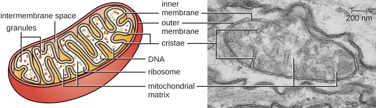
Image courtesy of CNX OpenStax
Nucleus
Appearance: Spherical or ovoid in shape, encased in a double membrane, the nuclear envelope, which is punctuated with pores.
Nuclear Pores: Protein complexes in the nuclear envelope that regulate the movement of molecules such as RNA and proteins between the nucleus and the cytoplasm.
Function: Repository of the cell’s DNA, thereby orchestrating cellular functions via gene expression.
Significance: Serves as the command centre of the cell, dictating protein synthesis and cellular replication.
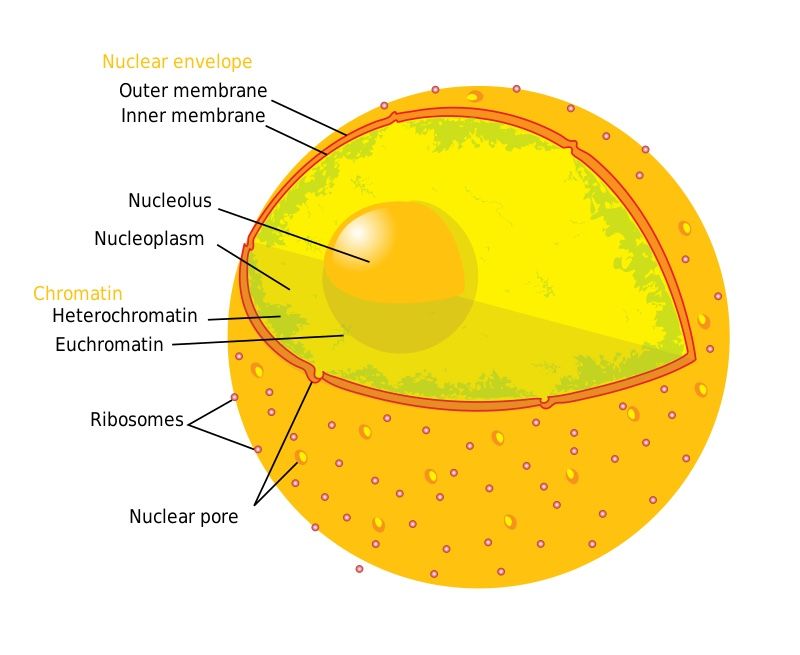
Image courtesy of Mariana Ruiz LadyofHats
Golgi Apparatus
Appearance: A series of closely stacked, flattened membranous sacs known as cisternae.
Function: It refines, warehouses, and ships cellular products, especially proteins and lipids.
Lipids: A diverse group of hydrophobic or amphipathic biological molecules, including fats, oils, and steroids, that play roles in energy storage, membrane structure, and signalling.
Significance: Plays a critical role in processing molecules synthesised in the cell and prepping them for transport.
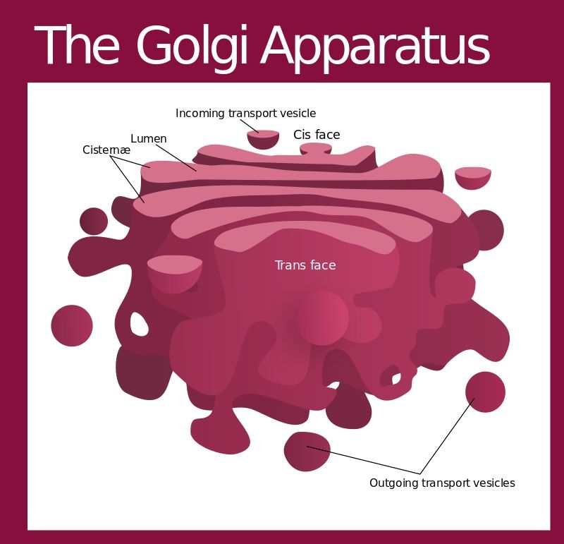
Image courtesy of Kelvinsong
Lysosome
Appearance: Membrane-bound vesicular structures brimming with digestive enzymes.
Function: They act as cellular waste disposal systems, breaking down unwanted materials.
Apoptosis: A form of programmed cell death in which a cell undergoes an orderly sequence of events leading to its elimination, critical for development and tissue homeostasis.
Significance: Beyond waste management, they play a role in cellular homeostasis and apoptosis (programmed cell death).
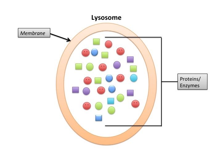
Image courtesy of lumoreno
Pitfalls and Remediation
Overcrowding: Overzealous labelling may cloud the main message. Prioritise key structures.
Omission of Functions: The omission of organelle function detracts from the understanding. Always incorporate it.
Scale Mismatch: A non-proportional drawing can lead to misinterpretations. Regularly cross-reference with the micrograph.
Mastery through Repetition
Regular Practice: The age-old adage "practice makes perfect" rings true. Regularly sketch different electron micrographs.
Feedback Loop: Solicit feedback. Peer or tutor evaluations can shed light on overlooked nuances.
Exam-centric Tips
Tool Preparedness: A sharp pencil and quality eraser are instrumental.
Anticipate Common Organelles: Familiarising oneself with commonly tested organelles can provide an edge.
Educated Guesses: In instances of uncertainty, deduce based on the organelle's appearance and its juxtaposition relative to other cellular structures.
Practice Questions
Drawing and annotating cell structures based on electron micrographs is pivotal in IB Biology for multiple reasons. First, the process reinforces learning by aiding visual memory, ensuring better retention of cellular details. Secondly, detailed and precise diagrams simplify the communication of intricate biological concepts. Lastly, it's an indispensable skill for examinations. The procedure involves selecting a clear, representative view of the structure, drawing with a sharp HB pencil while maintaining scale, and ensuring labels are succinct but informative. Including the function of each structure in the annotation offers a comprehensive understanding, allowing for effective communication of the cellular processes at play.
Upon receiving a diagram from an electron micrograph, my initial step would be to identify the organelle, ensuring a clear understanding of its appearance and function. I'd then replicate the organelle using a sharp HB pencil, meticulously maintaining the correct scale and proportions to prevent distortion. Continuous and smooth lines are essential for clarity. Labelling would be the subsequent step, where I'd draw straight lines towards the right side of the drawing, ensuring they don't overlap or clutter the diagram. Each label would be accompanied by a concise description and the function of the structure or organelle. Ensuring uniformity in font size and style would culminate in a neat, comprehensible representation.
FAQ
Maintaining correct scale and proportions requires meticulous observation and consistent referencing to the original electron micrograph. Students should start by outlining the primary boundaries or the general shape of the structure. Using grid paper or overlaying a transparent grid on the electron micrograph can help in ensuring proportional enlargement or reduction. It’s essential to frequently cross-reference between the drawing and the micrograph, ensuring every component is in proportion. Avoid rushing; taking the time to measure and compare distances and angles will go a long way in ensuring a proportional representation.
In IB Biology, colour annotations aren't typically required or recommended when working directly from electron micrographs, mainly because these micrographs are black and white and colour might introduce ambiguity. The priority is clarity and accuracy. However, using colour in study notes or for personal understanding can be beneficial as it can help distinguish between various structures or components. If students opt to use colour in personal notes, it's crucial to remain consistent and ensure that the colours don't overshadow or distort the original details of the micrograph.
Practice is essential for refining any skill. Students can source a variety of electron micrographs online or from textbooks. Regularly sketching these, and then comparing their drawings to the original image, is an excellent way to self-evaluate and improve. Joining study groups where peers can critique each other's work can provide fresh perspectives and highlight unnoticed mistakes. Additionally, there are many online resources and courses dedicated to scientific drawing, offering structured practice and expert feedback. Over time, consistent practice will build confidence and enhance the student's drawing and annotation skills.
Remembering functions can be challenging given the multitude of organelles. Mnemonics or memory aids can be incredibly effective. Creating a story or narrative that links various organelles and their functions can also assist memory. For example, visualising the endoplasmic reticulum as a factory conveyor belt, and the Golgi apparatus as the packaging department, can help remember their roles in protein synthesis and modification. Flashcards, with the organelle on one side and its function on the other, allow for repetitive testing and reinforcement. Over time, frequent revision and active recall make the information more accessible during annotations.
A sharp HB pencil is particularly recommended because it offers a balance between precision and visibility. The sharpness is crucial for reproducing intricate details seen in electron micrographs without making the lines too thick or rough. On the hardness scale, HB pencils are intermediate, meaning they provide dark enough lines to be clearly visible without being overly soft, which might smudge or create unwanted thick lines. Furthermore, the consistency of HB pencils ensures even, continuous lines, which are vital for presenting biological drawings with clarity and accuracy. They are also easily erasable, permitting corrections without leaving significant marks on the paper.
