
OCR Specification focus:
‘Describe xylem vessels, sieve tube elements and companion cells; examine and draw stained sections showing the distribution of xylem and phloem in organs.’
Vascular tissues in plants form an integrated transport network that supports water, mineral and assimilate movement. Their structure is closely related to efficient transport and mechanical support.
Structure and Function of Vascular Tissues
Overview of Plant Vascular Systems
Plants possess two main vascular tissues — xylem and phloem — organised into vascular bundles that extend through the roots, stems and leaves. These tissues transport essential substances and provide structural integrity. The precise arrangement within organs is crucial for transport efficiency and mechanical stability.
Xylem: Water and Mineral Transport
Composition and Structure
Xylem is responsible for the transport of water and dissolved mineral ions from the roots to aerial parts of the plant. It also provides mechanical support due to lignin deposition in its walls.
Xylem: A complex vascular tissue composed of dead cells specialised for the conduction of water and minerals and for structural support.
The main components of xylem include:
Xylem vessels – elongated, hollow tubes formed by the fusion of vessel elements end-to-end. The end walls are completely or partially removed to form a continuous column for water flow.
Tracheids – spindle-shaped cells with tapered ends and pits allowing lateral movement of water. These are more prominent in non-flowering plants.
Xylem parenchyma – living cells that store starch and facilitate lateral transport of water and minerals.
Fibres – elongated cells with thick lignified walls that add tensile strength.
Adaptations for Efficient Transport
Lignification strengthens vessel walls, preventing collapse under the tension created during transpiration.
Pits in vessel walls enable lateral movement of water between adjacent vessels.
Narrow lumens maintain capillary action for water ascent.
Continuous water columns facilitate cohesive water flow without obstruction.
Phloem: Translocation of Organic Solutes
Composition and Structure
Phloem is responsible for translocating organic molecules, particularly sucrose and other assimilates, from sources (photosynthetic tissues) to sinks (growing tissues, roots, storage organs).
Phloem: A complex living tissue responsible for the transport of organic solutes in plants through the process of translocation.
Phloem is composed of several cell types:
Sieve tube elements – elongated living cells that join end-to-end to form sieve tubes. The end walls contain sieve plates, perforated with pores that allow cytoplasmic continuity between cells.
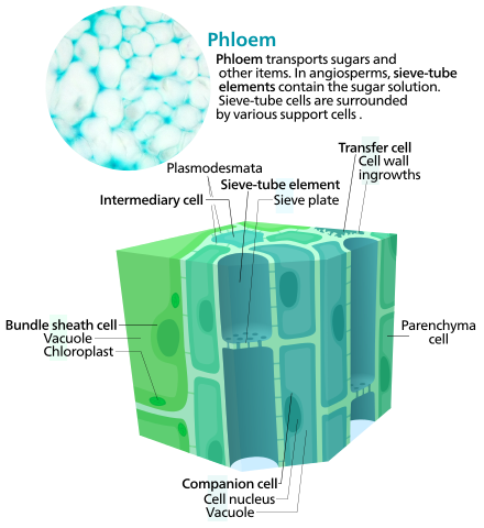
A labelled diagram of phloem showing a sieve tube element and its adjacent companion cell, connected by plasmodesmata. The sieve plate and reduced organelles in the sieve element illustrate how structure facilitates mass flow. This precisely matches the OCR requirement to describe sieve tubes and companion cells. Source.
Companion cells – metabolically active cells closely associated with sieve tubes, connected via plasmodesmata. They regulate metabolic functions and provide energy for active transport during loading and unloading of solutes.
Phloem parenchyma – involved in storage and lateral transport of nutrients.
Phloem fibres – provide additional structural support.
Functional Adaptations
Reduced cytoplasmic contents and absence of a nucleus in sieve elements allow unimpeded flow of phloem sap.
Mitochondria-rich companion cells actively pump sucrose into sieve tubes, maintaining concentration gradients for efficient transport.
Bidirectional flow within sieve tubes allows simultaneous transport to multiple sinks.
Organisation of Vascular Tissues in Plant Organs
Roots
In roots, vascular tissues are centrally located within the stele to resist pulling forces during water absorption.
Xylem forms a cross or star shape at the centre, with phloem occupying the spaces between the arms of the xylem.
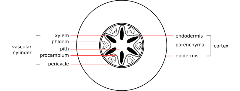
A schematic transverse section of a dicot root showing central xylem in a star-like pattern with phloem in between. The pericycle and endodermis surrounding the stele are shown to orient students when drawing. This reflects the typical organisation used to resist tensile forces during water uptake. Source.
Pericycle surrounds the vascular bundle and contributes to the formation of lateral roots.
Endodermis with the Casparian strip regulates the selective movement of water and solutes into the xylem.
This central arrangement maximises support against tension generated by transpiration pull.
Stems
In stems, vascular bundles are arranged in a ring near the periphery, supporting flexibility and upright growth.
Xylem is positioned towards the inside of each bundle, providing structural rigidity and water transport.
Phloem lies towards the outside, transporting assimilates from photosynthetic tissues.
The cambium, a layer of meristematic cells between xylem and phloem, allows secondary thickening through production of secondary vascular tissue.
This configuration provides both mechanical support and efficient transport pathways for long-distance movement.
Leaves
In leaves, vascular tissues form a branching network of veins essential for photosynthesis and transpiration.
The midrib contains the main vascular bundle, with smaller veins branching into the lamina.
Xylem occupies the upper side of the vein, ensuring a direct water supply to mesophyll cells for photosynthesis.
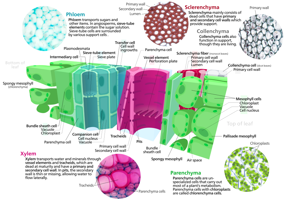
A high-quality leaf cross-section highlighting a vascular bundle with xylem on the upper side of the vein and phloem below. The figure also shows mesophyll and supporting tissues (sclerenchyma/collenchyma) — helpful but beyond the strict syllabus; students can ignore those labels if focusing only on vascular tissues. The clear labelling makes the positional relationship within the vein unambiguous. Source.
Phloem lies on the lower side, exporting photosynthetic products from the leaf to other parts of the plant.
This arrangement ensures close proximity between vascular tissues and mesophyll cells, maximising efficiency of gas exchange and transport.
Microscopic Observation of Vascular Tissues
Stained Sections
When viewing stained sections under a microscope:
Xylem stains brightly due to lignin, often appearing red or purple depending on the stain (e.g., toluidine blue or safranin).
Phloem typically stains a paler colour as its cells are living and non-lignified.
The pattern of vascular bundles helps identify the organ:
Root: star-shaped xylem at the centre.
Stem: vascular bundles arranged in a ring.
Leaf: network of veins with xylem uppermost.
Students should be able to identify and draw labelled diagrams of these structures, following OCR’s expectations for accurate biological drawings.
Functional Integration of Xylem and Phloem
Although xylem and phloem perform distinct roles, they are closely associated within vascular bundles, allowing coordination between water supply and assimilate distribution. The continuous vascular network ensures that water, minerals and organic solutes can move efficiently throughout the plant, maintaining physiological balance and supporting growth, photosynthesis, and reproduction.
Practice Questions
Question 1 (2 marks)
Describe two structural features of xylem vessels that enable them to transport water efficiently in plants.
Mark scheme:
Xylem vessels have no end walls / form a continuous column to allow unimpeded water flow (1 mark)
Their walls are thickened with lignin to prevent collapse under tension / provide mechanical support (1 mark)
(Other acceptable answers: narrow lumen to maintain capillary action; presence of pits for lateral movement of water — award 1 mark each, up to 2 total.)
Question 2 (5 marks)
Compare and contrast the structure and function of phloem sieve tube elements and companion cells.
Mark scheme:
Sieve tube elements: elongated cells joined end-to-end forming sieve tubes (1 mark)
Contain sieve plates with pores allowing cytoplasmic continuity between cells (1 mark)
Lack a nucleus and many organelles, allowing unimpeded flow of phloem sap (1 mark)
Companion cells: contain a nucleus and many mitochondria to provide energy for active transport processes (1 mark)
Connected to sieve tube elements by plasmodesmata for exchange of substances and coordination of activity (1 mark)
(Award full marks for correct comparison points; 1 mark per clear structural or functional distinction, up to 5 total.)
FAQ
Xylem vessels lose their cytoplasm and organelles as they mature, becoming hollow tubes. This reduces resistance to water flow and allows an unbroken column of water to move by cohesion and adhesion.
The lack of living contents also prevents metabolic activity from interfering with water transport and provides greater structural stability through lignified walls.
Companion cells use active transport to move sucrose into sieve tube elements against a concentration gradient.
Hydrogen ions are pumped out of the companion cell using ATP-driven proton pumps.
The resulting electrochemical gradient allows H+ ions to re-enter via co-transport proteins, bringing sucrose molecules in simultaneously.
Sucrose then diffuses into the sieve tube element through plasmodesmata.
This process maintains a high sucrose concentration in the phloem for translocation.
Different lignin deposition patterns allow flexibility or rigidity depending on plant part and function:
Annular or spiral patterns in young stems permit growth and elongation without breaking the vessels.
Reticulate or pitted patterns provide greater strength in mature tissues.
These patterns ensure that xylem remains both durable and adaptable to mechanical stresses and water pressure changes.
Roots position the vascular tissue centrally to resist pulling forces when absorbing water and anchoring the plant.
The xylem is central and star-shaped to withstand tension from the transpiration stream.
Phloem between the xylem arms allows close association for efficient nutrient exchange.
This compact arrangement also minimises damage from soil friction and supports lateral root growth from the pericycle.
In monocot stems, vascular bundles are scattered throughout the ground tissue with no defined ring. In dicot stems, they form a ring near the edge.
This difference affects secondary growth:
Dicots possess a vascular cambium between xylem and phloem, enabling thickening through secondary growth.
Monocots lack this cambium, so their stems generally remain slender and flexible.
The arrangement reflects each group’s typical growth habits and structural requirements.

.svg)