
OCR Specification focus:
‘Identify inner and outer mitochondrial membranes, cristae, matrix and mitochondrial DNA, relating structure to function.’
Mitochondria are specialised organelles that act as the cell’s powerhouses, efficiently converting chemical energy from nutrients into ATP through aerobic respiration. Their unique double-membrane structure and internal compartments enable efficient energy transfer, essential for maintaining life processes in eukaryotic organisms.
The Structure of the Mitochondrion
The Double Membrane System
Mitochondria are enclosed by two membranes: an outer mitochondrial membrane and an inner mitochondrial membrane, separated by an intermembrane space. Each membrane has distinct structural and functional properties that contribute to the organelle’s role in respiration.
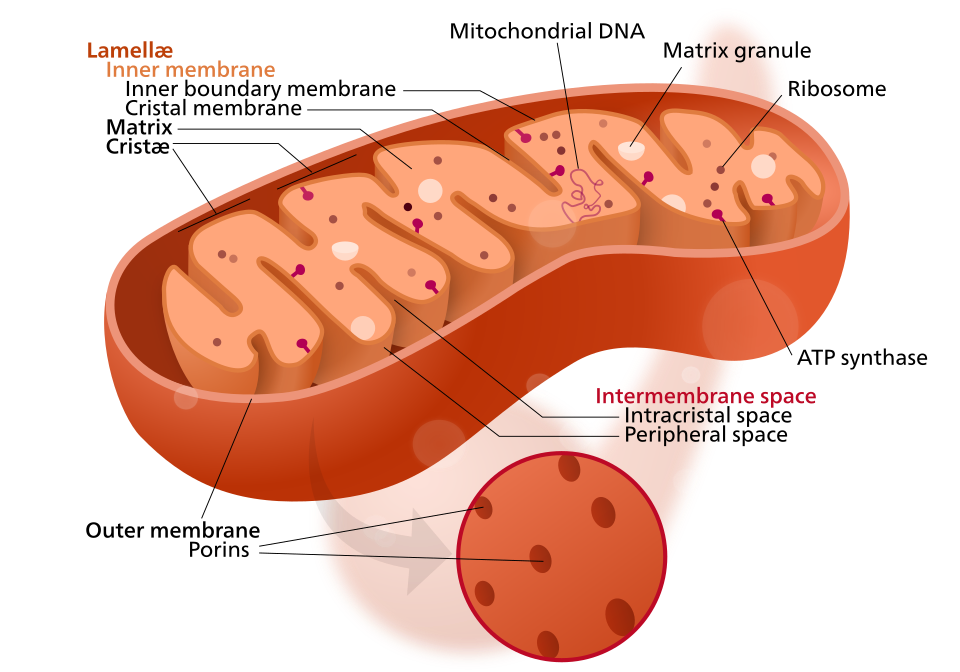
Diagram of a mitochondrion showing the outer membrane, inner membrane, cristae, intermembrane space, and matrix. The extensive folding of the inner membrane (cristae) greatly increases surface area for respiratory complexes. Labels are concise and match OCR terminology. Source.
Outer membrane
This smooth boundary contains porin proteins, which form large aqueous channels allowing free diffusion of ions, metabolites such as ATP and ADP, and small molecules up to around 10 kDa.
The outer membrane provides a protective barrier while maintaining permeability for essential substrates required for respiration.
Enzymes involved in lipid synthesis and the conversion of fatty acids are also embedded here.Inner membrane
This membrane is highly selectively permeable, controlling the passage of ions and molecules into the matrix. It houses the electron transport chain (ETC), ATP synthase, and transport proteins essential for oxidative phosphorylation.
Its folded nature increases surface area dramatically, enabling large numbers of enzyme complexes to be accommodated.
Cristae: Folds That Maximise Efficiency
Cristae are the inward folds of the inner mitochondrial membrane. Their main function is to increase surface area for the attachment of protein complexes involved in the final stages of respiration.
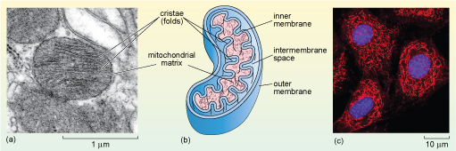
Electron micrograph of rat liver mitochondria with cristae projecting into the matrix and the double membrane visible at the boundary. This visual anchors the textbook diagram in authentic cell imagery. The page also shows a schematic and fluorescence image; those panels are extra context not required by the syllabus. Source.
The electron transport chain proteins and ATP synthase complexes are anchored on the cristae.
The degree of folding varies between cell types depending on energy demand; for instance, muscle cells contain mitochondria with densely packed cristae to maximise ATP production.
Cristae create micro-compartments that bring together substrates, enzymes, and coenzymes, improving reaction rates and energy efficiency.
The Mitochondrial Matrix
The matrix is the innermost compartment of the mitochondrion, enclosed by the inner membrane. It contains a complex mixture of enzymes, substrates, and cofactors necessary for the link reaction and the Krebs cycle.
Matrix: The internal fluid-filled space of the mitochondrion containing enzymes, mitochondrial DNA, ribosomes, and dissolved ions used for respiration.
Components of the Matrix
Enzymes catalyse reactions of the Krebs cycle and fatty acid oxidation.
Mitochondrial DNA (mtDNA) and ribosomes allow the synthesis of some mitochondrial proteins.
Metabolites such as pyruvate and coenzyme A are present for decarboxylation and dehydrogenation processes.
Ions and cofactors like magnesium (Mg²⁺) stabilise ATP and assist enzymatic activity.
The matrix serves as the site for CO₂ release and reduction of NAD⁺ and FAD, generating reduced coenzymes for use in oxidative phosphorylation on the inner membrane.
Mitochondrial DNA and Ribosomes
Mitochondria possess their own circular DNA molecule and 70S ribosomes, resembling those of prokaryotes.
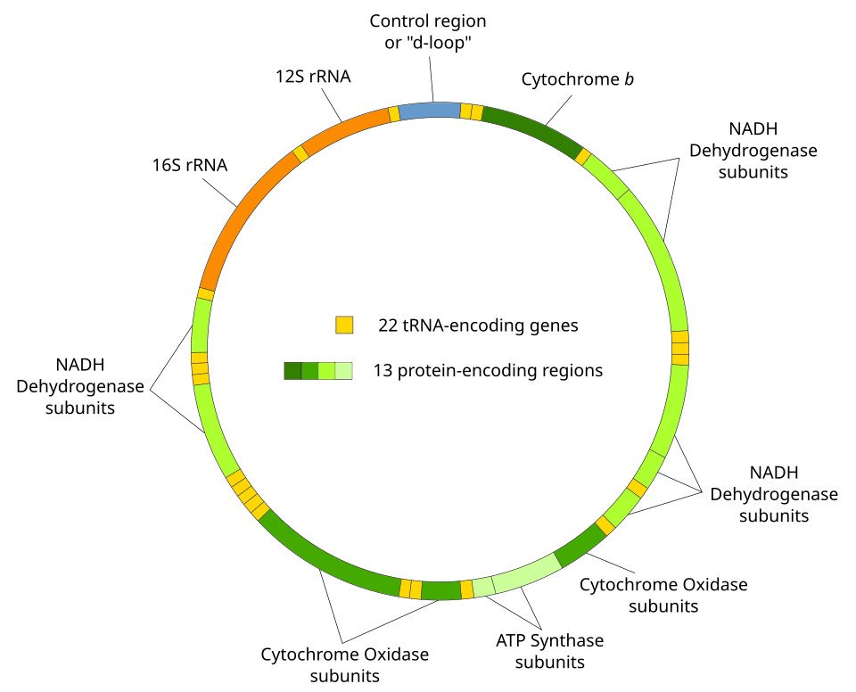
Circular map of human mitochondrial DNA (mtDNA), illustrating its compact genome organisation. Use this to emphasise that mtDNA is circular and housed in the matrix. Extra detail: individual gene names are shown; students do not need to memorise these for OCR. Source.
Mitochondrial DNA (mtDNA): A small, circular molecule of DNA within mitochondria that encodes some of the proteins and RNAs essential for mitochondrial function.
This genetic material supports the endosymbiotic theory, suggesting that mitochondria evolved from free-living bacteria engulfed by ancestral eukaryotic cells. The presence of mtDNA enables mitochondria to produce a limited number of proteins, tRNAs, and rRNAs independently of the nucleus. However, most mitochondrial proteins are still encoded by nuclear DNA and imported through specialised translocase complexes in the membranes.
The mitochondrial ribosomes synthesise essential inner membrane proteins, including components of the respiratory enzyme complexes. This partial genetic autonomy allows rapid adaptation to the cell’s energetic requirements.
The Intermembrane Space
The intermembrane space, between the inner and outer membranes, plays a vital role in proton accumulation during oxidative phosphorylation.
As electrons flow along the ETC, protons (H⁺ ions) are pumped into this space by complexes I, III, and IV of the inner membrane.
The resulting proton gradient forms a potential energy store, known as the proton motive force.
ATP synthase uses this gradient to drive ATP formation from ADP and inorganic phosphate when protons diffuse back into the matrix.
Thus, the compartmentalisation of the intermembrane space is crucial for maintaining energy coupling between redox reactions and phosphorylation.
Structural Adaptations and Functional Efficiency
Relationship Between Structure and Function
Each structural feature of the mitochondrion is precisely adapted to facilitate aerobic respiration and ATP synthesis:
The outer membrane regulates exchange and protects the organelle.
The inner membrane provides a platform for electron transport and chemiosmosis.
Cristae maximise the area for metabolic reactions.
The matrix hosts key metabolic cycles and contains genetic machinery.
The intermembrane space enables proton accumulation for ATP production.
Adaptations to Cellular Energy Demand
Cells with higher energy requirements contain mitochondria that are:
More numerous and larger in size.
Possessing densely folded cristae for increased surface area.
Found in regions of high metabolic activity, such as neurons and cardiac muscle cells.
Dynamic Nature of Mitochondria
Mitochondria are not static; they continuously move, divide, and fuse within the cytoplasm. These processes maintain a functional network capable of responding to energy fluctuations and stress conditions.
Mitochondrial fission allows distribution during cell division and removal of damaged organelles.
Fusion events help mix contents, including mtDNA, ensuring even distribution of genetic material and metabolic enzymes.
Their dynamic behaviour supports cellular homeostasis and efficient energy management.
Summary of Key Structural Features
Outer membrane: Permeable to small molecules; contains enzymes and porins.
Inner membrane: Selectively permeable; houses ETC and ATP synthase.
Cristae: Folds increasing surface area for ATP production.
Matrix: Site of Krebs cycle; contains mtDNA, enzymes, and ribosomes.
Intermembrane space: Reservoir for protons driving ATP synthesis.
mtDNA: Encodes essential proteins for mitochondrial metabolism.
Practice Questions
Question 1 (2 marks)
State two structural features of mitochondria that increase their efficiency in producing ATP, and explain how each feature contributes to this function.
Mark Scheme:
1 mark for identifying a correct structural feature.
1 mark for correctly linking that feature to its function.
Acceptable answers:
Cristae – increase surface area for attachment of enzymes and proteins involved in oxidative phosphorylation.
Inner membrane – contains electron transport chain and ATP synthase, enabling ATP production.
Matrix – contains enzymes for the Krebs cycle, generating reduced coenzymes for oxidative phosphorylation.
(Any two correct structure-function links, 1 mark each.)
Question 2 (5 marks)
Describe the structure of the mitochondrion and explain how its different components are adapted for their roles in respiration.
Mark Scheme:
Award marks for each relevant structural component correctly described and linked to its function.
1 mark – Description of the outer membrane as a smooth boundary containing porins that allow small molecules to pass while protecting the organelle.
1 mark – Description of the inner membrane as selectively permeable and containing proteins of the electron transport chain and ATP synthase complexes.
1 mark – Explanation of cristae as folds of the inner membrane that increase surface area for ATP production.
1 mark – Description of the matrix containing enzymes for the Krebs cycle, mitochondrial DNA, and ribosomes for protein synthesis.
1 mark – Reference to the intermembrane space as the site where protons accumulate to create a proton gradient driving ATP synthesis.
FAQ
The number and shape of mitochondria depend on the cell’s energy demands. Cells with high metabolic activity, such as muscle and sperm cells, contain more and larger mitochondria with densely packed cristae to increase ATP output.
In contrast, cells with lower energy requirements, like some connective tissue cells, have fewer mitochondria. Mitochondrial shape is dynamic — they can elongate or divide through fusion and fission processes to optimise energy distribution and remove damaged regions.
The outer membrane contains more phospholipids and fewer proteins, giving it greater permeability and flexibility. It includes porin channels that allow small molecules to pass.
The inner membrane, however, is rich in proteins (around 75% by mass) involved in the electron transport chain and transport systems. It lacks porins, making it highly selective. Its high protein-to-lipid ratio supports complex energy transduction processes.
Mitochondria retain DNA and ribosomes because they originated from an ancestral prokaryotic cell that formed a symbiotic relationship with a host cell.
Having their own genome allows them to synthesise a few essential proteins locally — mainly components of the respiratory complexes — ensuring rapid response to energy changes.
Most other mitochondrial proteins are encoded in nuclear DNA and imported via translocase systems, maintaining coordination between mitochondrial and cellular function.
Cristae structure adapts to the metabolic demands of the cell.
High-energy cells (e.g. cardiac muscle) have numerous, tightly packed cristae to maximise surface area for ATP production.
Lower-energy cells (e.g. adipose tissue) have fewer, simpler cristae.
Recent microscopy studies show that cristae can remodel dynamically, changing their density and curvature to match the cell’s activity level or stress conditions.
Structural defects — such as damaged cristae, faulty inner membrane proteins, or mutations in mitochondrial DNA — reduce the efficiency of oxidative phosphorylation.
Fewer or distorted cristae limit surface area for electron transport and ATP synthase activity.
Mutations in mtDNA can impair synthesis of key respiratory enzymes.
Damaged membranes may leak protons, disrupting the proton gradient.
Such defects can lead to mitochondrial diseases, fatigue, or reduced energy output in affected cells.
