
The nervous system allows the body to respond to internal and external stimuli through a complex network of cells, organs, and pathways that regulate all behavior.
The Nervous System
The nervous system is a highly complex, organized network responsible for detecting environmental changes, interpreting those changes, and responding accordingly. It controls virtually all body activities, from basic survival functions to the most advanced cognitive tasks. It relies on a system of specialized cells called neurons, which communicate through electrical impulses and chemical signals.
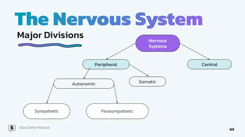
The nervous system is divided into two major parts:
The Central Nervous System (CNS), which consists of the brain and spinal cord, functions as the processing and command center.
The Peripheral Nervous System (PNS) includes all the nerves outside the CNS. It acts as a communication highway, carrying information between the CNS and the body’s limbs, organs, and tissues.
Both systems work in a coordinated manner to allow the human body to interact with its environment, maintain internal balance, and carry out tasks with precision.
Central Nervous System (CNS)
The central nervous system is the most critical control structure in the body. It receives incoming sensory information, interprets it, and responds by sending out instructions to various body parts. The CNS is protected by the skull (for the brain) and the vertebral column (for the spinal cord), as well as by the meninges (protective membranes) and cerebrospinal fluid (CSF), which cushions and nourishes the tissues.
The Brain
Although brain anatomy is addressed in another subtopic, its role within the CNS cannot be overlooked. The brain is the master control organ, responsible for both voluntary actions, like decision-making, and involuntary functions, such as maintaining heartbeat and respiration. It integrates sensory data, stores memories, creates emotions, and initiates behavior.
Primary Brain Functions within the CNS:
Integration of sensory information (sight, sound, touch, etc.)
Initiation of motor responses (movement)
Coordination of voluntary and involuntary activities
Regulation of internal environment (homeostasis)
Facilitation of cognition such as reasoning, language, and planning
The Spinal Cord
The spinal cord extends from the brainstem down the vertebral column and acts as the primary communication route between the brain and the rest of the body. It contains bundles of ascending nerve tracts (carrying sensory input to the brain) and descending tracts (carrying motor commands to the body).
The spinal cord also controls reflexes, which are rapid, automatic responses to stimuli. These do not require input from the brain and serve as protective mechanisms, such as the withdrawal reflex when touching something hot.
Functions of the Spinal Cord:
Transmitting sensory information from the PNS to the brain
Delivering motor commands from the brain to muscles and glands
Executing reflex actions independently from the brain
Peripheral Nervous System (PNS)
The peripheral nervous system connects the CNS to the limbs and organs. It is made up of cranial nerves, which emerge directly from the brain, and spinal nerves, which branch from the spinal cord. The PNS is subdivided into two major functional systems:
The Somatic Nervous System (SNS), which is under voluntary control
The Autonomic Nervous System (ANS), which regulates involuntary processes
The PNS includes both afferent (sensory) neurons and efferent (motor) neurons. Afferent neurons carry signals from sensory receptors toward the CNS, while efferent neurons transmit commands from the CNS to muscles or glands.
Primary Functions of the PNS:
Sensation: Detecting physical stimuli from the environment
Motor response: Delivering CNS output to muscles for movement
Communication: Transmitting data to ensure responsiveness and coordination
Somatic Nervous System (SNS)
The somatic nervous system governs all voluntary muscular activity and is involved in processing sensory information that comes from external sources. This includes information such as temperature, light, sound, and touch.
Components of the SNS:
Motor neurons: Extend from the CNS to skeletal muscles to control movement
Sensory neurons: Transmit information about stimuli from the skin, muscles, and joints to the CNS
Key Features:
Voluntary control over movement, posture, and coordination
Conscious perception of stimuli such as pain and pressure
Involves a single neuron in the motor pathway from CNS to muscle
Enables quick, intentional responses to environmental changes
Somatic Reflexes
Reflexes mediated by the somatic nervous system involve quick, involuntary responses. These are controlled by reflex arcs, which are neural pathways that bypass the brain for speed.
Example: The patellar reflex (knee-jerk response) occurs when the patellar tendon is tapped, and a signal is rapidly sent through the spinal cord, resulting in immediate leg extension.
Autonomic Nervous System (ANS)
The autonomic nervous system oversees involuntary body functions such as heart rate, digestion, respiratory rate, pupil dilation, and glandular activity. It maintains homeostasis by controlling internal organs without conscious effort.
The ANS is divided into:
The sympathetic nervous system
The parasympathetic nervous system
Each division innervates the same organs but produces opposite effects to maintain physiological balance.
Sympathetic Nervous System
The sympathetic division is responsible for the body’s "fight or flight" response, which is activated during stress, danger, or intense activity.
Effects of Sympathetic Activation:
Increased heart rate and respiration
Dilation of pupils
Increased blood flow to muscles
Reduced activity of the digestive system
Release of glucose from liver for energy
Stimulation of sweat glands
This system is activated by norepinephrine and epinephrine (adrenaline), which are released by sympathetic nerve endings and the adrenal medulla.
Parasympathetic Nervous System
The parasympathetic division supports "rest and digest" functions. It promotes relaxation, recovery, and maintenance of bodily resources after stress has passed.
Effects of Parasympathetic Activation:
Decreased heart rate
Constriction of pupils
Stimulation of digestion and nutrient absorption
Relaxation of urinary bladder
Energy conservation
This system operates through the neurotransmitter acetylcholine, which slows down physiological activity and restores balance.
Dual Innervation
Most target organs are controlled by both sympathetic and parasympathetic nerves. This dual innervation provides dynamic control. For example:
In the heart:
Sympathetic nerves increase heart rate
Parasympathetic nerves decrease heart rate
In the digestive tract:
Sympathetic nerves suppress digestion
Parasympathetic nerves promote digestion
This counterbalance allows the body to swiftly adapt to changing internal or external conditions.
Enteric Nervous System (ENS)
The enteric nervous system is sometimes referred to as the “second brain” because it operates largely independently from the CNS. It is responsible for regulating the digestive system, including:
Movement of food through the gastrointestinal tract (peristalsis)
Enzyme secretion
Blood flow to the digestive organs
Although it can function independently, the ENS is influenced by both sympathetic and parasympathetic divisions. For example, stress can suppress digestion via sympathetic activation, while a relaxed state enhances digestion via parasympathetic control.
Key Characteristics:
Contains approximately 100 million neurons, more than the spinal cord
Found within the walls of the esophagus, stomach, intestines, and colon
Communicates with the CNS through the vagus nerve
Uses neurotransmitters like serotonin and dopamine, many of which are also found in the brain
Comparing Somatic and Autonomic Nervous Systems
Somatic Nervous System:
Voluntary control
Connects CNS to skeletal muscle
Single neuron from CNS to target
Always excitatory (activates muscle)
Autonomic Nervous System:
Involuntary control
Connects CNS to smooth muscle, cardiac muscle, and glands
Two-neuron chain: preganglionic and postganglionic
Can be excitatory or inhibitory
Involves dual innervation
Nervous System Coordination
Everyday bodily responses rely on integration between the CNS and PNS. For example:
If you hear an alarm clock:
Sensory receptors in your ears detect the sound (PNS).
The signal travels via afferent neurons to your brain (CNS).
The brain processes the sound and determines you need to wake up.
A signal is sent via efferent neurons to skeletal muscles (SNS) to move.
Simultaneously, sympathetic activation increases heart rate and alertness (ANS).
This highly coordinated interaction allows for fluid, real-time behavioral and physiological responses.
Recap of Divisions and Their Functions
CNS: Brain and spinal cord; decision-making, processing, and command initiation.
PNS: Sensory and motor nerves outside the CNS; communication between body and brain.
SNS: Controls voluntary movement and skeletal muscle activity.
ANS: Regulates involuntary functions; split into:
Sympathetic: Fight-or-flight, arousal.
Parasympathetic: Rest-and-digest, relaxation.
ENS: Manages digestion; functions semi-independently.
The integration and cooperation of these systems ensure that organisms can survive, adapt, and interact with their surroundings effectively. The complexity and adaptability of the nervous system make it one of the most essential biological systems in the body.
FAQ
Although all action potentials have the same electrical strength due to the all-or-nothing principle, the nervous system differentiates between weak and strong stimuli by frequency of firing and number of neurons activated. Stronger stimuli cause:
Increased frequency of action potentials in individual neurons (higher rate of firing).
Recruitment of additional sensory neurons, meaning more neurons are stimulated simultaneously.
Spatial summation, where input from multiple neurons is processed together.
This coding allows the brain to interpret intensity without changing the amplitude of each signal, ensuring consistent and accurate communication regardless of stimulus strength.
Glial cells, or neuroglia, are non-neuronal support cells that play crucial roles in maintaining nervous system function. They do not conduct impulses but are essential for healthy signaling. Key roles include:
Astrocytes: Regulate the extracellular environment, maintain the blood-brain barrier, and provide nutrients to neurons.
Oligodendrocytes (CNS) and Schwann cells (PNS): Form the myelin sheath that insulates axons and speeds up impulse transmission.
Microglia: Act as immune cells, removing pathogens and debris through phagocytosis.
Ependymal cells: Produce and circulate cerebrospinal fluid.
Glial cells are vital for protecting, nourishing, and structurally supporting neurons throughout the nervous system.
The nervous system ensures unidirectional flow of information by the specific structure and function of neurons and synapses:
Signals always travel from dendrites to axons due to the neuron’s design.
Axon terminals release neurotransmitters into the synaptic cleft.
Post-synaptic receptors on the next neuron detect and respond to these neurotransmitters.
Voltage-gated ion channels only open in one direction, ensuring forward propagation.
Refractory periods prevent a second action potential from firing backward.
This setup guarantees that nerve impulses travel efficiently and in one direction from sensory input to processing centers to motor output.
The effects of spinal cord damage depend on the location and severity of the injury. Since the spinal cord contains both ascending (sensory) and descending (motor) tracts:
Cervical damage (neck region) may result in quadriplegia, affecting both arms and legs, and possibly breathing if high enough.
Thoracic damage can impair trunk and leg functions but typically spares the arms.
Lumbar or sacral damage usually affects lower body movement and bladder control.
Damage to one side of the spinal cord may lead to loss of motor function on the same side and loss of pain/temperature sensation on the opposite side (Brown-Séquard syndrome).
The specific effects align with the structure and function of affected spinal tracts.
Cranial and spinal nerves both belong to the peripheral nervous system, but they differ in several important ways:
Cranial nerves (12 pairs) originate directly from the brain or brainstem, while spinal nerves (31 pairs) emerge from the spinal cord.
Cranial nerves mostly control functions of the head and neck, including vision, hearing, taste, facial expression, and some autonomic control (e.g., heart rate via the vagus nerve).
Spinal nerves serve the rest of the body, providing both sensory input and motor output to limbs, torso, and organs.
Some cranial nerves are sensory only, some are motor only, and others are mixed. In contrast, all spinal nerves are mixed nerves, containing both sensory and motor fibers.
These differences reflect their specialized roles in coordinating regional body functions.
Practice Questions
Explain how the sympathetic and parasympathetic divisions of the autonomic nervous system work together to maintain homeostasis in the human body.
The sympathetic and parasympathetic divisions of the autonomic nervous system maintain homeostasis by exerting opposite but complementary effects on target organs. The sympathetic division activates the "fight-or-flight" response during stress by increasing heart rate, dilating airways, and inhibiting digestion. In contrast, the parasympathetic division promotes the "rest-and-digest" state by slowing the heart rate, constricting airways, and stimulating digestive activity. Both systems constantly adjust internal functions based on environmental demands. This dual innervation ensures rapid responses to change while allowing recovery and balance, enabling the body to maintain a stable internal environment essential for survival and optimal function.
Describe the differences in structure and function between the somatic and autonomic divisions of the peripheral nervous system.
The somatic and autonomic divisions differ in both structure and function. The somatic system controls voluntary movements using one motor neuron to transmit signals directly from the central nervous system to skeletal muscles. It enables conscious control and responses to external stimuli. In contrast, the autonomic system regulates involuntary functions such as heart rate and digestion using a two-neuron chain: a preganglionic neuron and a postganglionic neuron. It operates without conscious control and is further divided into sympathetic and parasympathetic branches. These differences allow the body to perform both deliberate actions and automatic regulation efficiently and simultaneously.
