
IB Syllabus focus: 'Regulation of the heart depends on intrinsic and extrinsic excitation. The heart's structure is shown in the SEHS data booklet and supports understanding of cardiac control.'
The heart regulates its rhythm through built-in pacemaker tissue and outside nerve influences. Understanding how these systems interact explains coordinated contraction, changing heart rate, and effective blood movement during rest and activity.
Intrinsic excitation
The heart’s built-in pacemaker system
The heart has an intrinsic ability to generate its own electrical impulse. This is why the heart is described as myogenic: the stimulus to contract begins in the cardiac muscle tissue itself rather than requiring a nerve to trigger each beat.
Intrinsic excitation: Cardiac activation generated from within the heart by specialized pacemaker and conducting tissue.
This intrinsic system is made of specialized cardiac cells that repeatedly produce and transmit electrical signals. Other parts of the conduction system can also generate impulses, but the normal rhythm is set by the region that fires fastest.
The key starting point is the sinoatrial (SA) node, located in the wall of the right atrium. The SA node is the heart’s natural pacemaker because its cells spontaneously depolarize at a regular rate. Since it normally fires faster than other possible pacemaker regions, it controls the rhythm of the whole heart. Each impulse spreads across the atrial walls, causing the two atria to contract.
The impulse next reaches the atrioventricular (AV) node. The AV node briefly delays transmission. This delay is essential because it gives the atria time to empty blood into the ventricles before the ventricles contract. If this delay did not occur, the pumping sequence would be less effective.
After the AV node, the impulse travels down the bundle of His in the septum and then through the Purkinje fibers in the ventricular walls. This pathway causes the ventricles to contract in a coordinated way, beginning near the apex and moving upward so blood is pushed effectively out of the heart.
Intrinsic excitation is responsible for:
initiating each heartbeat
setting the heart’s basic rhythm
coordinating atrial contraction before ventricular contraction
producing an efficient, ordered pumping action
Heart structure and the conduction pathway
Why the route of excitation matters
The structure of the heart supports its electrical control. The atria and ventricles have different roles, so they must contract in the correct sequence. If they contracted at the same time, ventricular filling would be reduced and cardiac pumping would become less effective.
Because the SA node, AV node, bundle of His, and Purkinje fibers are found in specific locations, the excitation wave follows an organized route instead of spreading randomly. In the SEHS data booklet, identifying the right atrium, septum, and ventricular walls helps you trace this route of control.
The septum is especially important because it carries conducting tissue toward the ventricles. This helps both ventricles contract in a coordinated pattern rather than as separate, poorly timed chambers.
The sequence is:
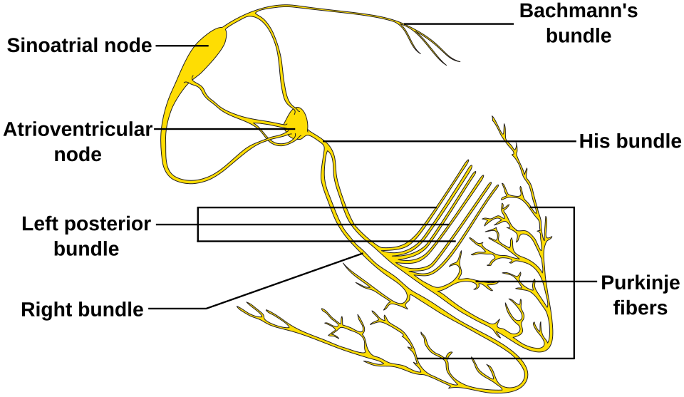
Diagram of the heart’s electrical conduction system showing the anatomical locations of the SA node, AV node, bundle of His (AV bundle), and Purkinje fibers. This visual helps you connect the route of excitation to the timing of atrial contraction before ventricular contraction. Source
SA node fires
impulse spreads across both atria
atria contract
impulse reaches the AV node
a short delay occurs
impulse travels through the bundle of His
impulse spreads through the Purkinje fibers
ventricles contract
This conduction pathway allows the heart to act as a coordinated pump rather than as separate pieces of muscle. If the pathway is disrupted, timing becomes less efficient and the heart’s pumping action can be reduced.
Extrinsic excitation
Outside influences on cardiac control
Although the heart can beat independently, it does not always beat at the same rate. Signals from outside the heart adjust the intrinsic pacemaker system so the circulation matches the body’s needs.
Extrinsic excitation: Regulation of cardiac activity by signals originating outside the heart, mainly autonomic nerve input.
Extrinsic excitation does not usually start each heartbeat from nothing. Instead, it modifies the rate and pattern already created by the intrinsic system. These outside influences mainly affect the SA node, AV node, and cardiac muscle, altering timing and force without replacing the normal conduction route.
The main external control comes from the autonomic nervous system:
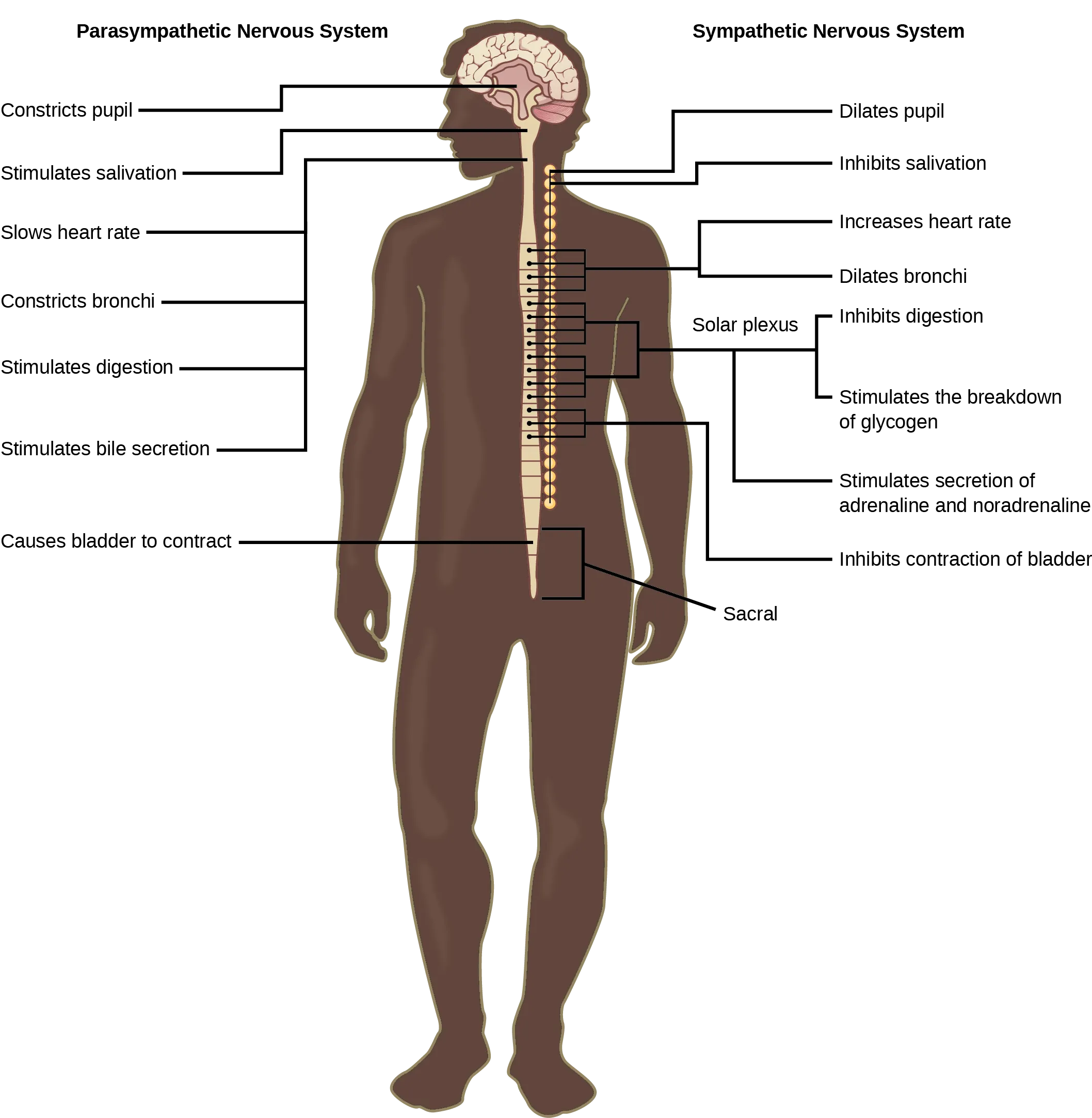
Overview diagram contrasting sympathetic (“fight-or-flight”) and parasympathetic (“rest-and-digest”) divisions of the autonomic nervous system and their target-organ effects. For the heart, it reinforces the key exam idea that sympathetic input increases heart rate, while parasympathetic (vagal) input slows it. Source
Sympathetic stimulation increases the firing rate of the SA node, speeds conduction through the heart, and increases the force of contraction.
Parasympathetic stimulation, mainly through the vagus nerve, slows the firing rate of the SA node and slows conduction through the AV node.
At rest, parasympathetic influence is usually greater, which helps keep heart rate lower than the SA node’s own intrinsic pace. When demand increases, such as at the beginning of exercise, parasympathetic influence is withdrawn and sympathetic input rises. This allows the heart to beat faster and pump more effectively.
Extrinsic control is important because it provides flexibility. The intrinsic system gives the heart a dependable rhythm, while the extrinsic system adjusts that rhythm according to current physiological demand.
Intrinsic and extrinsic excitation working together
The most important idea is that intrinsic excitation creates the heartbeat, while extrinsic excitation regulates that heartbeat. In a healthy heart, this regulation is continuous from beat to beat.
The intrinsic system provides:
pacemaker activity from the SA node
conduction through the AV node, bundle of His, and Purkinje fibers
the correct order of atrial and ventricular contraction
The extrinsic system provides:
faster or slower heart rate
changes in conduction speed
changes in contraction strength when needed
This interaction can be seen across normal daily conditions:
during quiet rest, parasympathetic activity slows pacemaker firing
at the onset of activity, reduced parasympathetic influence allows heart rate to rise
as activity continues, sympathetic stimulation further increases cardiac response
during recovery, parasympathetic influence becomes more dominant again
The heart therefore does not depend on a nerve to make every beat happen, but it does depend on outside input to match its activity to the body’s needs. Understanding both forms of excitation helps explain how heart structure, electrical conduction, and rate control combine to maintain effective circulation.
Practice Questions
State the roles of the SA node and the AV node in intrinsic excitation of the heart. [2]
SA node acts as the natural pacemaker / initiates the electrical impulse / starts atrial contraction. [1]
AV node delays and relays the impulse to the ventricles / allows ventricles to contract after the atria. [1]
Explain how intrinsic and extrinsic excitation work together to regulate the heart when a person moves from rest to exercise. [5]
Intrinsic excitation means the heart can generate its own impulse because it is myogenic. [1]
SA node initiates the impulse and sets the basic rhythm. [1]
The impulse passes through the AV node and then through the bundle of His and Purkinje fibers to coordinate ventricular contraction. [1]
As exercise begins, parasympathetic influence decreases and/or sympathetic influence increases. [1]
This increases SA node firing rate and speeds cardiac activity, so the heart responds to the greater demand. [1]
FAQ
A transplanted heart can keep beating because the SA node and the rest of the conduction system are intrinsically myogenic. They do not need a direct nerve impulse to start each heartbeat.
However, loss of normal nerve connections means early control of heart rate is less precise. Resting heart rate is often higher, and changes in rate can be slower until some limited reinnervation occurs.
If the SA node fails, other parts of the conduction system may take over as secondary pacemakers. The AV node is the usual backup, but it fires at a slower rate.
This can lead to a slower heartbeat and less efficient rhythm control. If the resulting rhythm is too slow or unstable, an artificial pacemaker may be needed to maintain normal cardiac timing.
The AV node delay gives the atria time to finish contracting before the ventricles begin. This improves ventricular filling.
Without that delay, the atria and ventricles would contract too close together. The ventricles would receive less blood before pumping, reducing the effectiveness of each heartbeat.
An artificial pacemaker replaces or supports a faulty intrinsic pacemaker or conduction pathway. It sends timed electrical impulses so the heart contracts in a more normal pattern.
Some pacemakers are also rate-responsive. That means they can increase pacing during movement, partly mimicking the adjustments that would normally occur through extrinsic control.
The anatomical conduction pathway is usually the same, but training changes how it is regulated. Endurance athletes typically have greater parasympathetic tone at rest.
They also tend to eject more blood per beat, so the heart does not need to beat as often to maintain circulation at rest. The lower resting rate is therefore mainly a regulation difference, not a structural change in the conduction route.
