

Question 1
A participant's heart rate was measured during recovery immediately after exercise. Which interpretation best integrates the graph with autonomic nervous-system control?
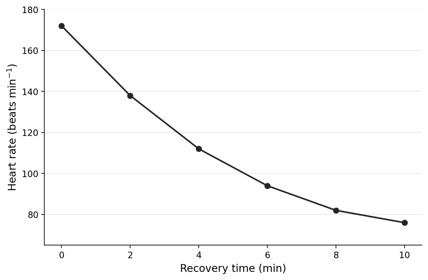
A. Sympathetic influence becomes increasingly dominant so that heart rate returns towards its exercise value.
B. Parasympathetic influence becomes greater as heart rate returns towards its pre-exercise condition.
C. The somatic nervous system secretes insulin to produce the fall in heart rate.
D. Chemoreceptors increase core temperature so that heart rate decreases.
Question 2
A participant's core temperature rose following an external thermal challenge and then began to stabilise. Which pair of responses is most consistent with negative-feedback thermoregulation at the elevated temperatures shown?
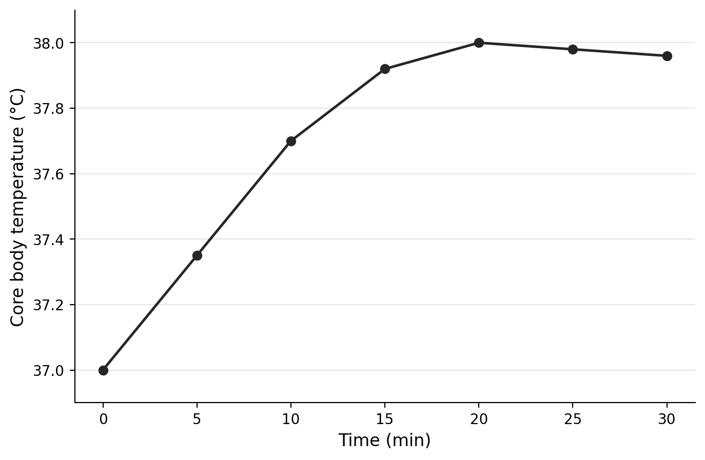
A. Vasoconstriction and shivering
B. Vasoconstriction and non-shivering thermogenesis
C. Shivering and non-shivering thermogenesis
D. Sweating and vasodilation
Question 3
A student recorded heart rate while one participant completed exercise at progressively greater intensities. The nervous system coordinates cardiac responses, including through the autonomic nervous system.
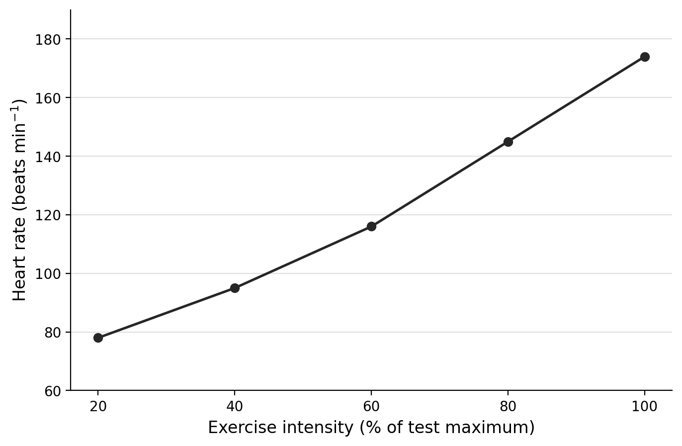
Describe the relationship between exercise intensity and heart rate. Calculate the change in heart rate between and of test maximum.
Explain how the sympathetic and parasympathetic divisions of the autonomic nervous system could contribute to the pattern shown as exercise intensity increases.
Predict the direction of change in heart rate during recovery after exercise and explain your prediction using the concept of physiological regulation.
Question 4
In a hypothetical investigation, a brief challenge caused arterial blood pressure to rise before it gradually returned towards its initial value. Baroreceptors are among the specialised receptors that respond to stimuli and initiate nervous-system responses.
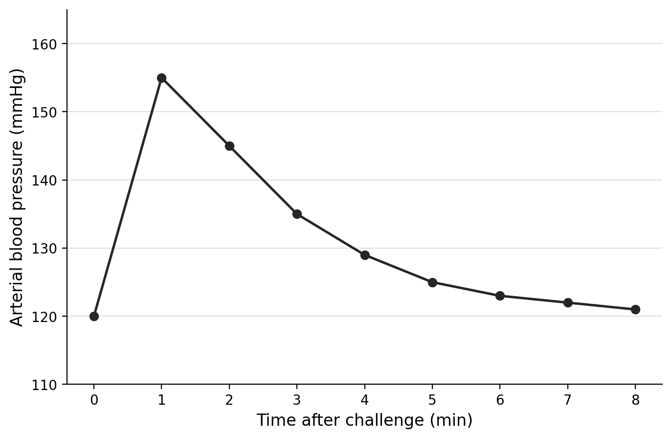
Describe the change in arterial blood pressure after the challenge.
Identify the specialised receptor most directly associated with detecting a change in blood pressure and outline its role in the response.
Explain how the graph provides evidence of negative feedback and homeostasis.
Question 5
The diagram below is a simplified representation of communication between the central nervous system and the internal and external environments. Treat the arrows as showing sensory information towards the central nervous system and motor information away from it.
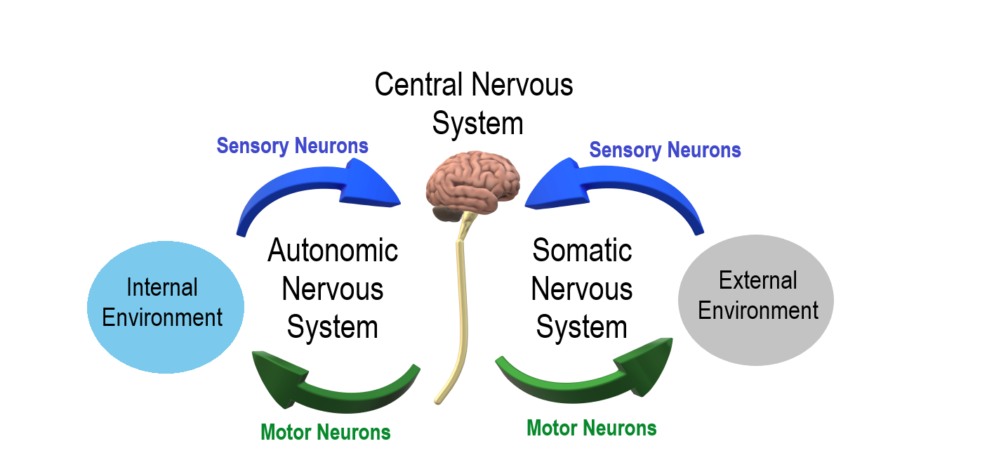
Using the diagram, distinguish between the autonomic and somatic nervous systems in terms of their association with the internal and external environments.
State the two principal divisions of the nervous system.
State how the efferent division is subdivided, and then state the two subdivisions of the autonomic nervous system.
An athlete changes from rest to exercise. Explain how the communication pattern represented in the diagram can enable the nervous system to coordinate cardiac function, breathing and ventilation, and temperature control as conditions change.
Question 6
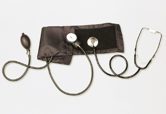
The photograph below shows conventional mechanical equipment used to measure a participant's blood pressure before and after exercise.
Name the specialised receptor type from A.1 Communication that is most directly associated with a blood-pressure stimulus.
A change in blood pressure is recorded using the equipment shown. Explain how detection of this change can be connected with nervous-system regulation of cardiac function. Detailed internal functioning of the receptor is not required.
A student concludes that a change in blood pressure measured after exercise is sufficient to describe the complete cardiovascular response to the exercise. Evaluate this conclusion using other cardiovascular variables from A.1 Communication.
Question 7
The photograph below shows a runner during sustained outdoor exercise. A visible skin response provides evidence that thermoregulation is active.
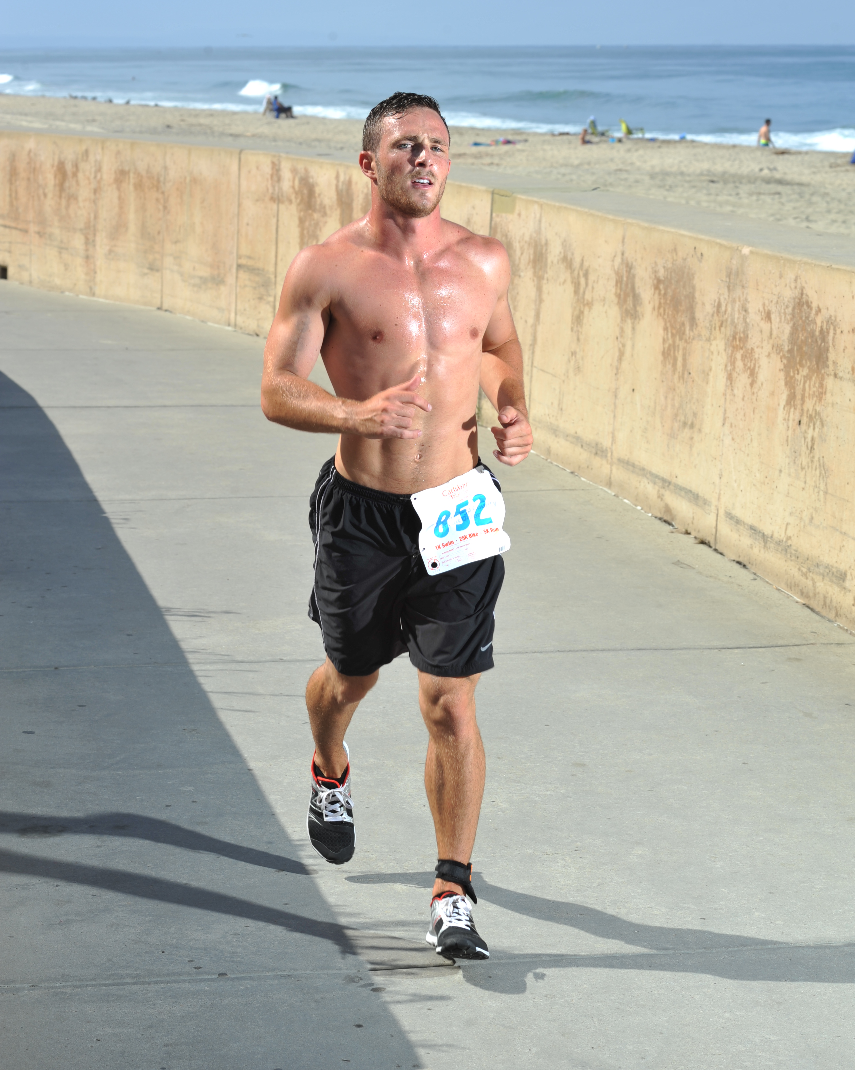
Identify the thermoregulatory response that is visibly evident in the photograph. State one other thermoregulatory response that could operate when core body temperature requires regulation during exercise.
Explain how the visible response can form part of negative feedback when core body temperature moves away from approximately .
Two athletes perform the same activity in the same external environment but show different thermoregulatory responses. Analyse this observation using three factors identified in A.1 Communication.
Question 8
The photograph below shows a glucometer used to obtain repeated measurements from a participant during an SEHS investigation.
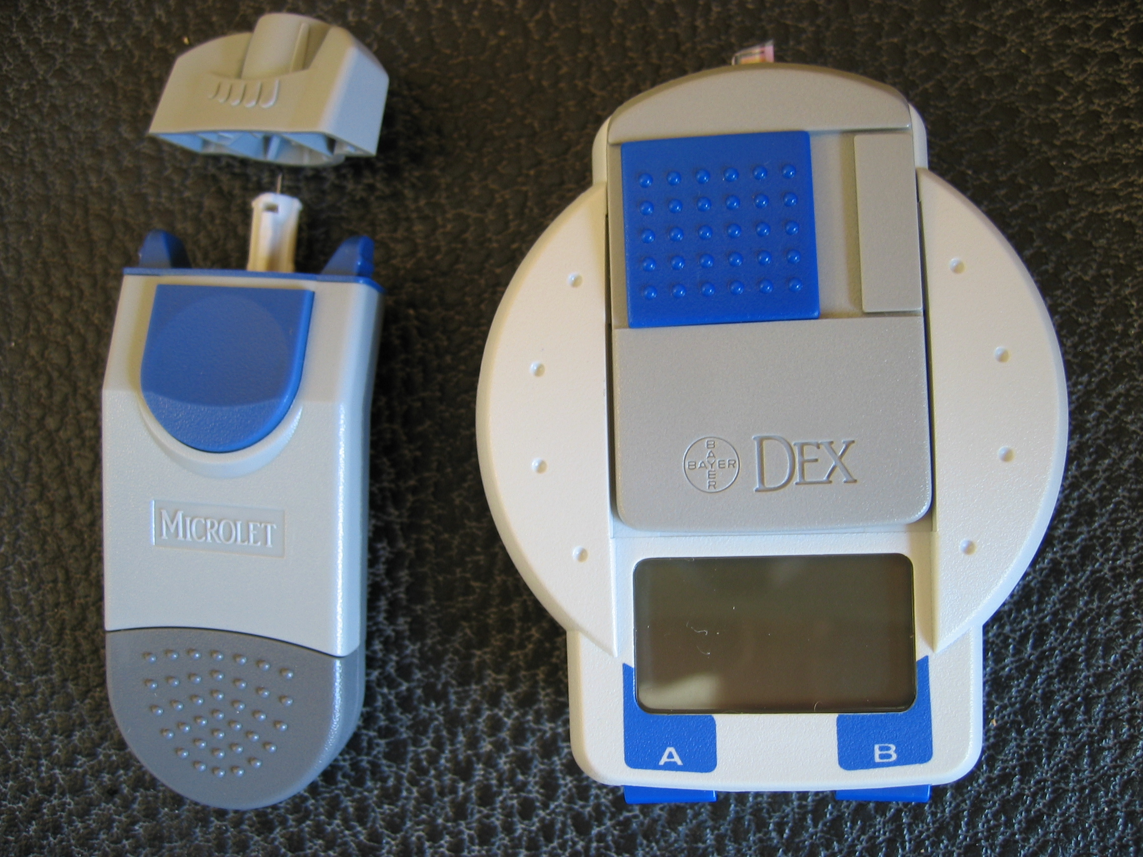
State the homeostatically regulated variable monitored using the equipment shown and name the two hormones identified in A.1 Communication that regulate this variable.
Explain how regulation of this variable illustrates the principle of negative feedback. Detailed biochemical mechanisms are not required.
Measurements are taken before and during exercise. Explain the two effects of exercise on blood-glucose regulation that are explicitly identified in A.1 Communication.
Question 9
Which statement correctly describes the organization of the nervous system?
A. The central nervous system is a subdivision of the peripheral nervous system.
B. The somatic nervous system is a subdivision of the central nervous system.
C. The nervous system is divided into the central nervous system and peripheral nervous system.
D. The peripheral nervous system is divided only into sympathetic and parasympathetic nervous systems.
Question 10
The labelled diagram below shows the extensive distribution of major blood vessels around the human body. Do not infer physiological meaning from colour alone; use the visible distribution and labels as evidence.
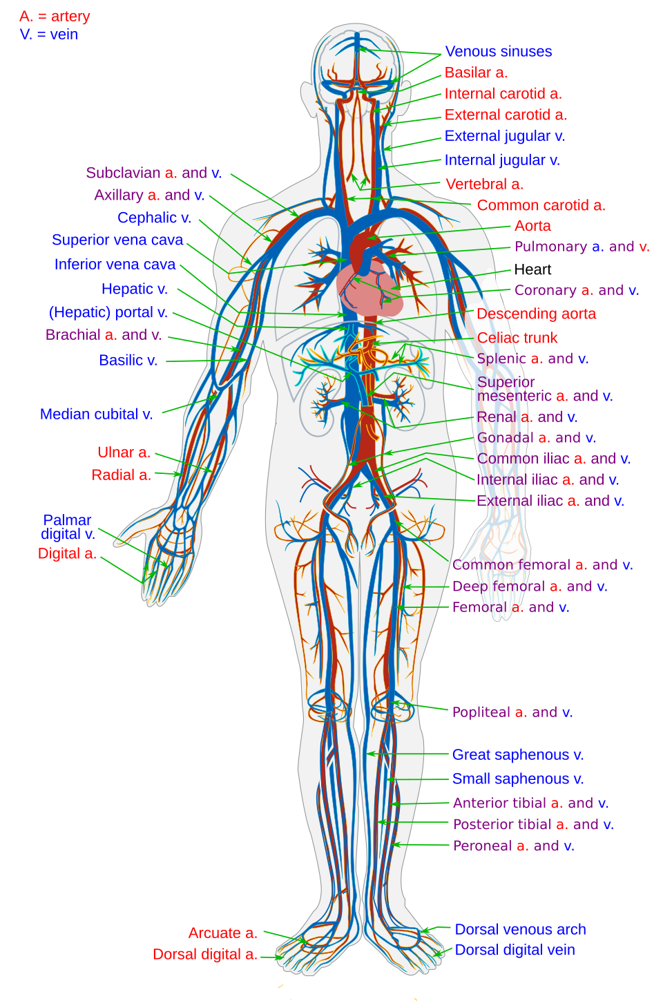
Using the diagram, describe two visible features that demonstrate the body-wide distribution of the cardiovascular system.
Explain how the widespread distribution visible in the image supports the transport functions of the cardiovascular system. Include four substances or materials identified in A.1 Communication.
A student claims that the static diagram proves that cardiovascular activity remains unchanged between low- and high-intensity exercise. Evaluate this claim.