
OCR Specification focus:
‘Describe phospholipids, cholesterol, proteins, glycoproteins and receptors where hormones and drugs bind.’
The fluid mosaic model describes the dynamic structure of cell membranes, combining lipids, proteins, and carbohydrates to form selectively permeable boundaries essential for communication and regulation.
The Fluid Mosaic Model
The fluid mosaic model proposes that the cell surface membrane (plasma membrane) consists of a phospholipid bilayer with embedded proteins, cholesterol, and carbohydrates. The components move laterally, giving the membrane flexibility and fluidity while maintaining structural integrity. This model explains how membranes can function as selectively permeable barriers, sites of communication, and reaction surfaces.
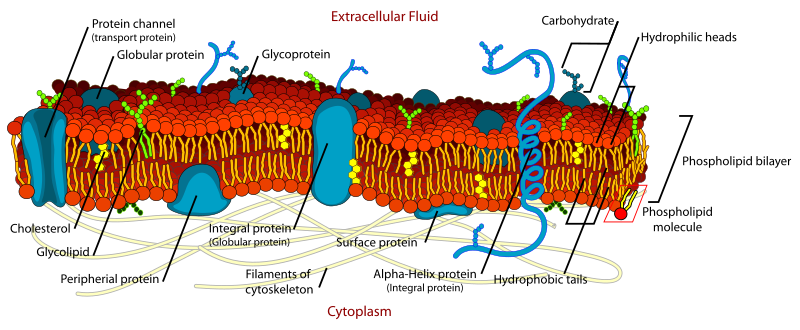
Labeled diagram of the fluid mosaic membrane, highlighting the phospholipid bilayer with cholesterol interspersed, and intrinsic/extrinsic proteins forming channels and carriers. Glycoproteins and glycolipids extend into the extracellular space to mediate cell recognition and signalling. The clean layout aligns with OCR-level detail without excess information. Source.
Phospholipid Bilayer
Phospholipids form the fundamental structure of biological membranes. Each molecule has a hydrophilic phosphate head and hydrophobic fatty acid tails, resulting in a bilayer arrangement where:
Hydrophilic heads face outward towards the aqueous environments inside and outside the cell.
Hydrophobic tails face inward, creating a non-polar interior that prevents free passage of water-soluble substances.
Phospholipid bilayer: A double layer of phospholipid molecules forming the structural basis of cell membranes, with hydrophilic heads outward and hydrophobic tails inward.
This arrangement allows small non-polar molecules (e.g. oxygen, carbon dioxide) to diffuse freely while restricting ions and polar molecules, maintaining membrane selectivity.
Membrane Fluidity and Cholesterol
Cholesterol, a lipid with a rigid ring structure, is interspersed among phospholipids and plays a key role in regulating membrane fluidity:
At high temperatures, cholesterol prevents excessive movement of phospholipids, maintaining stability.
At low temperatures, it prevents phospholipids from packing too closely, maintaining flexibility.
This regulation ensures membrane integrity and supports cell survival in varying conditions.
Cholesterol: A lipid molecule that modulates membrane fluidity and stability by fitting between phospholipids within the bilayer.
Cholesterol also reduces membrane permeability to small polar molecules, reinforcing the selective barrier.
Membrane Proteins: Intrinsic and Extrinsic
Proteins are vital functional components of the membrane. They contribute to transport, cell signalling, enzyme activity, and cell recognition.
Intrinsic (Integral) Proteins
These span the entire bilayer and often form channels or carriers:
Channel proteins create hydrophilic pores allowing specific ions or molecules to pass by facilitated diffusion.
Carrier proteins bind molecules and change shape to transport them across, sometimes using ATP in active transport.
Intrinsic protein: A membrane protein embedded within the phospholipid bilayer that facilitates transport or acts as a receptor.
Extrinsic (Peripheral) Proteins
These are located on one side of the membrane, loosely attached by electrostatic interactions or hydrogen bonds. They often act as enzymes, anchors, or cell identifiers.
Glycoproteins and Glycolipids: Recognition and Communication
Glycoproteins (proteins with attached carbohydrate chains) and glycolipids (lipids with carbohydrate chains) extend from the membrane surface into the extracellular space, forming the glycocalyx — a carbohydrate-rich zone critical for cell recognition and signalling.
Key functions include:
Acting as antigens for immune recognition (e.g. ABO blood groups).
Serving as receptors for hormones, neurotransmitters, and drugs.
Enabling cell adhesion in tissue formation.
Protecting cells from mechanical and chemical damage.
Glycoprotein: A protein with carbohydrate chains attached, involved in cell recognition and communication.
Receptors and Cell Signalling
Cell membranes are not passive barriers; they actively mediate communication between the cell and its environment.
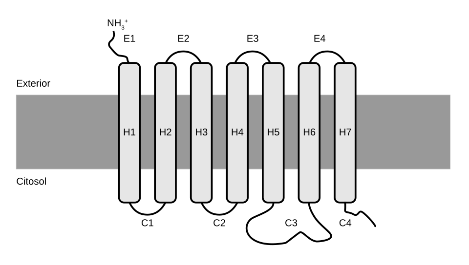
Schematic of a G-protein–coupled receptor (GPCR) spanning the membrane with an extracellular ligand-binding site and intracellular G-protein interaction. This illustrates how a receptor protein detects a chemical signal and initiates signal transduction. Extra detail (the heterotrimeric G-protein components) is depicted but not required beyond recognising that receptors trigger intracellular responses. Source.
Receptor proteins embedded in the membrane detect chemical signals (ligands) such as hormones, neurotransmitters, or drugs.
Ligand-Receptor Interactions
When a ligand binds to its specific receptor:
The receptor changes shape (conformational change).
This triggers a cascade of intracellular events (a signal transduction pathway).
The cell responds — for example, by altering gene expression or activating an enzyme.
Receptor: A specific protein on a cell membrane or within a cell that binds a signalling molecule to elicit a response.
This process underpins cell communication and homeostasis, allowing coordination across tissues and organs.
Examples
Insulin receptors on muscle and liver cells bind insulin, triggering glucose uptake.
Adrenaline receptors activate second messenger systems that stimulate metabolic responses.
Drug receptors, such as those for beta-blockers, can mimic or inhibit natural ligands to regulate physiological processes.
Mosaic Nature of the Membrane
The term “mosaic” reflects the patchwork of proteins, lipids, and carbohydrates within the bilayer. Each component performs specialised roles:
Structural proteins maintain cell shape.
Transport proteins control movement of substances.
Receptor and recognition molecules manage communication and identity.
Enzymes catalyse reactions at the membrane surface.
Together, these create a dynamic, adaptable structure vital for cellular function.
Importance of the Fluid Mosaic Structure
The fluid mosaic arrangement ensures membranes are:
Flexible, allowing cells to change shape during movement or division.
Self-sealing, enabling vesicle formation and fusion in endocytosis and exocytosis.
Selective, controlling entry and exit of materials.
Responsive, with receptors enabling cells to detect and respond to environmental changes.
Biological membranes, therefore, are integral to maintaining cell integrity, communication, and metabolic coordination.
Practice Questions
Question 1 (2 marks)
Describe the role of cholesterol in the phospholipid bilayer of cell membranes.
Mark Scheme:
1 mark for stating that cholesterol regulates membrane fluidity.
1 mark for stating that cholesterol prevents membranes from becoming too rigid at low temperatures or too fluid at high temperatures (accept “stabilises the membrane”).
Question 2 (5 marks)
Explain how the fluid mosaic structure of the cell surface membrane enables it to perform its functions in transport and cell communication.
Mark Scheme:
Award up to 5 marks for the following indicative content (maximum 1 mark per valid point):
Phospholipid bilayer forms a selectively permeable barrier, allowing non-polar molecules to diffuse while preventing passage of polar molecules and ions.
Intrinsic (integral) proteins form channels and carriers that enable facilitated diffusion and active transport across the membrane.
Cholesterol within the bilayer maintains membrane fluidity and stability, ensuring correct function of embedded proteins.
Glycoproteins and glycolipids act in cell recognition and signalling, forming part of the glycocalyx.
Receptor proteins on the surface bind hormones or neurotransmitters, triggering intracellular responses (signal transduction).
FAQ
Membrane fluidity influences how easily proteins move and interact within the bilayer. If the membrane becomes too rigid, proteins may not change shape or migrate efficiently to carry out transport or signalling. Conversely, excessive fluidity can disrupt the correct alignment of protein channels and receptors.
Maintaining an optimal fluid state ensures:
Efficient transport via carrier and channel proteins
Proper receptor–ligand binding and signal transduction
Stable yet flexible cell structure
Each side of the membrane differs in composition and function. The outer layer is rich in glycolipids and glycoproteins, forming the glycocalyx for recognition and signalling, whereas the inner layer contains more phospholipids and peripheral proteins involved in anchoring and metabolism.
This asymmetry is maintained during vesicle formation, ensuring each membrane retains its orientation when moving through the cell.
Unsaturated fatty acids have kinks due to double bonds, preventing phospholipids from packing tightly. This increases membrane fluidity and permeability.
More unsaturated fatty acids = more fluid membranes
More saturated fatty acids = more rigid membranes
Cells can adjust fatty acid composition to adapt to temperature changes, ensuring consistent membrane behaviour across different environments.
A faulty receptor may:
Fail to bind its ligand (e.g. hormone or neurotransmitter)
Bind but not trigger signal transduction
Send continuous, uncontrolled signals
This can lead to cellular dysfunction or disease. For example, defective insulin receptors prevent glucose uptake, contributing to insulin resistance in type 2 diabetes. Some cancers also involve overactive receptor signalling that drives uncontrolled cell growth.
The term “mosaic” refers to the patchwork arrangement of proteins, lipids, and carbohydrates in the membrane. Each component performs distinct yet coordinated functions:
Phospholipids form the structural foundation
Proteins enable transport, catalysis, and signalling
Carbohydrates provide identity and recognition
This diverse organisation allows membranes to be both functional and adaptable, constantly rearranging without losing integrity.
