
OCR Specification focus:
‘Sensory receptors, such as Pacinian corpuscles, detect specific stimuli and act as transducers, converting stimulus energy to nerve impulses.’
Sensory receptors detect and respond to environmental stimuli, converting diverse forms of energy into electrical nerve impulses that can be interpreted by the nervous system.
The Role of Sensory Receptors
Sensory receptors are specialised cells or nerve endings that detect changes in the internal or external environment and initiate nervous responses. They are essential for survival, allowing organisms to detect stimuli such as light, temperature, pressure, and chemical changes.
External stimuli include light, sound, touch, and temperature changes.
Internal stimuli include blood pressure, pH, and solute concentration.
Characteristics of Sensory Receptors
Specificity – each receptor type responds to one particular form of stimulus (e.g. photoreceptors detect light).
Transduction – receptors convert the energy of the detected stimulus into an electrical signal.
Adaptation – sensitivity of receptors can change if a stimulus is constant, allowing the organism to focus on new changes.
Threshold – a minimum level of stimulus intensity is required to trigger a response and generate an action potential.
Transducer: A cell or organ that converts one form of energy into another, specifically converting stimulus energy into electrical energy in the form of nerve impulses.
Types of Sensory Receptors
Each receptor type detects a specific form of energy and is found in different parts of the body:
Photoreceptors – respond to light energy, located in the retina of the eye.
Chemoreceptors – detect chemical stimuli, such as oxygen or carbon dioxide levels in blood.
Thermoreceptors – respond to temperature changes, located in the skin and hypothalamus.
Mechanoreceptors – detect mechanical pressure or movement, for example in the skin or inner ear.
Nociceptors – respond to painful stimuli resulting from damage to tissue.
Each of these receptors ensures precise detection and appropriate neural signalling for different types of stimuli.
The Pacinian Corpuscle as an Example
The Pacinian corpuscle is a well-studied example of a mechanoreceptor found deep in the skin, particularly in areas such as the fingers, soles of feet, and joints. It detects mechanical pressure and vibrations.
Structure
A Pacinian corpuscle consists of:
A sensory nerve ending at its centre.
Surrounding concentric layers of connective tissue (lamellae) separated by viscous gel.
A capsule enclosing the corpuscle and insulating it from other stimuli.
When pressure is applied to the skin, the lamellae become deformed, transmitting mechanical energy to the nerve ending.
Sensory receptors, such as Pacinian corpuscles, detect specific stimuli and act as transducers, converting stimulus energy into nerve impulses.
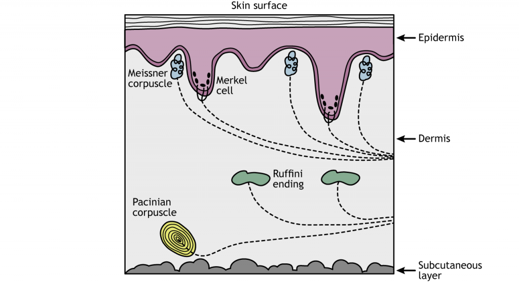
Illustrated cross-section showing the positions and functions of the skin’s mechanoreceptors, including Pacinian corpuscles deep in the dermis for vibration and deep pressure. The diagram highlights receptor depth and receptive roles without unnecessary detail. Labels are concise and appropriate for A-level study. Source.
Generation of a Nerve Impulse
The process by which a receptor converts stimulus energy into an electrical impulse is called transduction. In the Pacinian corpuscle, this involves mechanically gated sodium channels and changes in membrane potential.
Steps in Transduction within a Pacinian Corpuscle
Resting State:
The membrane of the sensory neurone is polarised.
Sodium ion (Na⁺) channels are closed.
The inside of the cell is negatively charged relative to the outside.
Application of Pressure:
The lamellae deform, causing the stretch-mediated sodium channels to open.
Sodium ions diffuse into the neurone.
Generator Potential Formation:
The influx of Na⁺ causes depolarisation of the membrane.
If the depolarisation reaches the threshold potential, an action potential is triggered.
Action Potential Transmission:
The action potential travels along the sensory neurone to the central nervous system, carrying information about the intensity and duration of the stimulus.
In a Pacinian corpuscle, deformation of the lamellated capsule opens mechanosensitive Na⁺ channels in the neurone ending, initiating a graded generator potential.
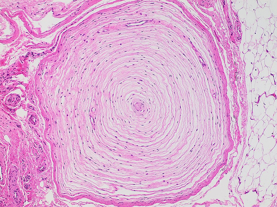
Light micrograph of a Pacinian corpuscle in the dermis, displaying the concentric lamellae that mechanically filter sustained pressure yet transmit rapid vibrations to the nerve ending. This encapsulation underlies rapid adaptation and effective transduction of vibratory stimuli into nerve impulses. The image shows histological detail only and does not add content beyond the syllabus. Source.
Generator potential: A small, local change in electrical potential across the receptor cell membrane caused by a stimulus. It can trigger an action potential if a threshold is reached.
Features of the Generator Potential
Graded response – the magnitude of depolarisation depends on the stimulus intensity.
If the threshold is not reached, the potential dissipates without triggering an impulse.
Multiple weak stimuli in quick succession can combine (summation) to reach threshold and generate an action potential.
This ensures that only sufficiently strong or persistent stimuli are transmitted to the brain.
Action potential: A rapid, temporary reversal of the electrical potential difference across a neurone membrane when a nerve impulse is transmitted.
Frequency Coding of Stimulus Intensity
Although each action potential is of the same amplitude, the frequency of action potentials varies with stimulus intensity:
Weak stimuli produce low-frequency impulses.
Strong stimuli generate high-frequency impulses.
This allows the brain to interpret stimulus strength based on the rate of impulses arriving from sensory receptors.
Adaptation of Receptors
Some receptors exhibit sensory adaptation, reducing their response to a constant stimulus over time. The Pacinian corpuscle, for example, responds strongly when pressure is first applied or removed but not to continuous pressure. This mechanism helps the nervous system focus on new or changing stimuli, rather than unchanging background information.
Integration and Role in the Nervous System
Sensory receptors act as the interface between the environment and the nervous system, initiating the sequence of neuronal communication:
Detection – receptors sense specific stimuli.
Transduction – conversion of stimulus energy to electrical energy.
Transmission – action potential travels via sensory neurone to the CNS.
Interpretation – CNS processes the information to produce an appropriate response.
The accurate detection and conversion of stimuli underpin an organism’s ability to maintain homeostasis and respond appropriately to environmental changes.
Key Summary Points
Sensory receptors are specialised transducers that detect specific stimuli and generate electrical impulses.
The Pacinian corpuscle exemplifies mechanoreception through deformation-induced depolarisation.
Generator potentials are graded; only reaching the threshold produces an action potential.
Frequency of impulses encodes stimulus strength, enabling the brain to interpret environmental information.
Receptors such as photoreceptors, chemoreceptors, thermoreceptors, and mechanoreceptors each have distinct roles in stimulus detection and neural signalling.
Practice Questions
Question 1 (2 marks)
Describe the role of the Pacinian corpuscle in detecting mechanical stimuli.
Mark scheme:
1 mark: States that the Pacinian corpuscle detects mechanical pressure or vibrations.
1 mark: Explains that deformation of the lamellae opens stretch-mediated sodium channels, resulting in depolarisation or a generator potential.
Question 2 (5 marks)
Explain how a Pacinian corpuscle acts as a transducer to convert mechanical pressure into a nerve impulse.
Mark scheme:
1 mark: Mentions that mechanical pressure deforms the lamellae of the Pacinian corpuscle.
1 mark: States that this deformation causes stretch-mediated sodium channels in the sensory neurone membrane to open.
1 mark: Describes sodium ions (Na⁺) diffusing into the neurone, causing depolarisation.
1 mark: Explains that if the depolarisation reaches a threshold potential, an action potential is generated.
1 mark: Notes that the action potential is then transmitted along the sensory neurone to the central nervous system (CNS).
FAQ
The Pacinian corpuscle is rapidly adapting because its concentric lamellae are separated by fluid, allowing the structure to quickly redistribute pressure. This means that the receptor only responds when pressure is applied or released, not when it is constant.
As a result, it is particularly effective at detecting vibrations and changes in mechanical force, rather than continuous touch. This rapid adaptation allows the nervous system to prioritise new sensory information.
Pacinian corpuscles are found deep in the dermis of the skin, as well as in joint capsules, tendons, and ligaments.
Their deep placement enables them to detect deep pressure and vibrations transmitted through tissues rather than light touch.
In fingertips, they provide fine tactile discrimination.
In joints, they help detect movement and changes in joint angle, contributing to proprioception — the awareness of body position.
The generation of an action potential requires sufficient influx of sodium ions to make the inside of the membrane positive relative to the outside.
If the stimulus is too weak, only a small depolarisation (generator potential) occurs, and voltage-gated sodium channels remain closed.
Only when the membrane reaches the threshold potential do these channels open, triggering the full all-or-nothing response characteristic of action potentials.
Generator potentials are graded, meaning their magnitude depends on the strength of the stimulus. They occur locally at the receptor membrane and can summate if stimuli are repeated quickly.
In contrast, action potentials are non-graded — they occur fully or not at all once the threshold is reached. They are propagated along the neurone without loss of amplitude, allowing reliable transmission of information to the CNS.
Pacinian corpuscles are mechanoreceptors, so they only respond to mechanical pressure or vibration.
They cannot detect:
Thermal changes, as they lack thermoreceptive ion channels.
Chemical stimuli, because they have no receptors for specific molecules.
Light, as they are not connected to phototransduction pathways.
Their selective sensitivity ensures efficient sensory coding, allowing other receptors (e.g. thermoreceptors, chemoreceptors) to handle different environmental cues.
