
OCR Specification focus:
‘Explain how the resting potential is established and maintained and how an action potential is generated, including positive feedback.’
Nerve impulses transmit information rapidly along neurones, using precisely controlled changes in membrane potential. Understanding resting potentials and action potentials reveals how electrical signals underpin nervous communication.
Resting Potential: Establishment and Maintenance
At rest, a neurone maintains an electrical potential difference across its plasma membrane of approximately –70 mV, with the inside being negative relative to the outside.
Ionic Basis of Resting Potential
This resting potential results from unequal distribution of sodium (Na⁺) and potassium (K⁺) ions, controlled by membrane permeability and active transport.
The phospholipid bilayer is impermeable to ions; therefore, channel proteins and pumps control ion movement.
The sodium–potassium pump (Na⁺/K⁺-ATPase) actively transports 3 Na⁺ ions out and 2 K⁺ ions in per ATP molecule hydrolysed.
The membrane is more permeable to K⁺ due to open potassium leak channels, allowing K⁺ to diffuse out down its concentration gradient.
Fewer Na⁺ ions diffuse in because fewer Na⁺ channels are open at rest.
The result is a net loss of positive charge from the neurone’s interior, creating a negative resting potential.
Resting potential: The steady electrical charge difference across the neurone membrane when no impulse is being transmitted, typically about –70 mV.
Role of the Sodium–Potassium Pump
The pump maintains the ionic gradients essential for excitability:
Uses ATP to transport ions against their concentration gradients.
Ensures Na⁺ remains high outside and K⁺ high inside.
Without this, the electrochemical gradient would dissipate, preventing action potential generation.
Action Potential: Generation and Transmission
When a neurone is stimulated sufficiently, the membrane potential changes rapidly and temporarily becomes positive inside. This is the action potential—a short, self-propagating electrical impulse.
Threshold and Depolarisation
A stimulus must cause the membrane potential to reach the threshold (about –55 mV) to trigger an action potential.
Depolarisation begins when voltage-gated sodium channels open.
Na⁺ ions rush in by diffusion, following both concentration and electrical gradients.
The membrane potential becomes positive, peaking at around +30 mV.
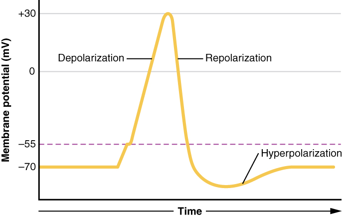
Graph of a neurone’s membrane potential over time, illustrating threshold, depolarisation, repolarisation, and after-hyperpolarisation. Values approximate –70 mV at rest and +30 mV at peak, matching typical mammalian neurones. Labels focus on the phases required by the specification. Source.
Depolarisation: The reduction and reversal of the resting membrane potential due to the influx of sodium ions.
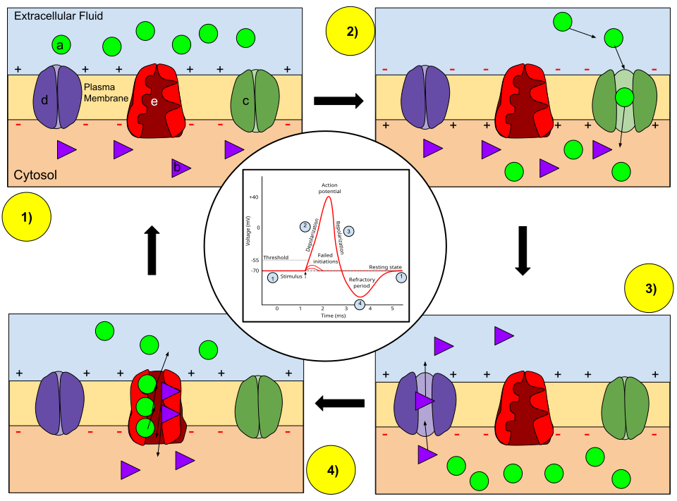
The rapid Na⁺ influx is driven by positive feedback:
The initial depolarisation opens more voltage-gated Na⁺ channels.
Increased Na⁺ entry further depolarises the membrane.
This continues until all sodium channels are open.
Annotated diagram showing resting state, depolarisation (Na⁺ channels open), repolarisation (K⁺ channels open), and return to rest, with the Na⁺/K⁺-ATPase maintaining gradients. Channel states are clearly indicated; legend includes extra reference to intracellular anions beyond the OCR emphasis. Source.
Positive Feedback in Action Potentials
Positive feedback ensures an all-or-nothing response:
If the threshold is reached, the response is full-scale and uniform.
If not reached, no action potential occurs.
The strength of stimulus affects frequency, not amplitude, of impulses.
Repolarisation and Hyperpolarisation
After depolarisation, the neurone must restore resting conditions.
Repolarisation
At +30 mV, voltage-gated Na⁺ channels close.
Voltage-gated K⁺ channels open, and K⁺ diffuses out of the cell.
This returns the inside to negative relative to the outside.
Repolarisation: The restoration of negative membrane potential as potassium ions leave the axon following depolarisation.
Hyperpolarisation
The outward diffusion of K⁺ briefly overshoots the resting potential.
The membrane potential may fall to –80 mV before stabilising.
This phase prevents immediate re-excitation.
Refractory Period
Following an action potential, the membrane enters a refractory period during which another impulse cannot be generated:
Ensures unidirectional propagation of impulses.
Limits maximum impulse frequency, allowing discrete signal coding.
Ionic Movements and Membrane Permeability Changes
Throughout an action potential:
Resting state: Na⁺ and K⁺ channels closed (except leak channels).
Depolarisation: Na⁺ channels open; Na⁺ influx dominates.
Repolarisation: K⁺ channels open; K⁺ efflux dominates.
Recovery: Sodium–potassium pump restores ion gradients.
The cycle lasts about 2–5 milliseconds, enabling rapid signalling along axons.
The All-or-Nothing Principle
Action potentials do not vary in size. Once threshold potential is reached:
The depolarisation always reaches approximately +30 mV.
A stronger stimulus increases impulse frequency, not magnitude.
This coding method allows the nervous system to represent stimulus intensity.
Propagation of the Action Potential
Local Currents
Depolarisation at one region of the membrane causes Na⁺ ions to diffuse sideways within the axoplasm:
These local currents depolarise the adjacent section of the membrane.
Voltage-gated Na⁺ channels in that region open, propagating the action potential.
Saltatory Conduction
In myelinated neurones, myelin sheaths insulate axons, preventing ion movement except at nodes of Ranvier:
Action potentials jump from node to node (saltatory conduction).
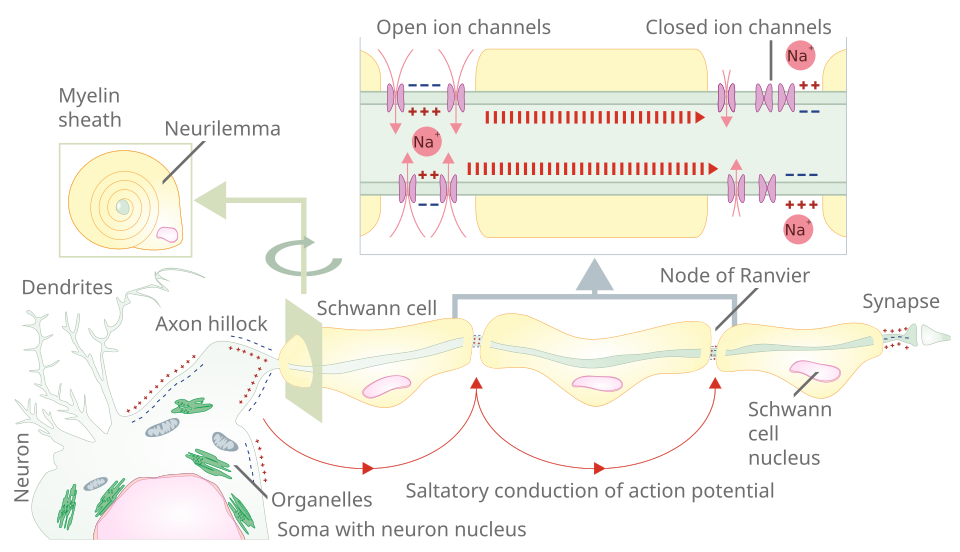
Schematic of a myelinated axon demonstrating saltatory conduction: action potentials regenerate at nodes of Ranvier while myelin insulates internodes to increase speed. Labels indicate Schwann cells, nodes, and the direction of propagation; includes peripheral myelin terminology consistent with OCR language. Source.
Greatly increases speed of transmission, up to 100 m s⁻¹.
Saltatory conduction: The leaping transmission of action potentials between nodes of Ranvier in a myelinated axon.
Unmyelinated neurones conduct impulses continuously, with slower overall transmission.
Recovery and Restoration
Once the action potential passes:
Na⁺ channels reset to closed but responsive.
K⁺ channels close gradually.
The Na⁺/K⁺ pump and leak channels re-establish the resting ionic balance.
EQUATION
—-----------------------------------------------------------------
Membrane Potential (Vₘ) = V_inside – V_outside
Vₘ = Potential difference across the membrane (millivolts, mV)
V_inside = Electrical potential of cytoplasm
V_outside = Electrical potential of extracellular fluid
—-----------------------------------------------------------------
This equation defines the basis of neuronal excitability, determining when depolarisation can initiate a new impulse.
Summary of Key Mechanisms
Resting potential: maintained by Na⁺/K⁺ pumps and K⁺ leak channels.
Depolarisation: Na⁺ influx via voltage-gated channels.
Positive feedback: amplifies depolarisation to action potential.
Repolarisation: K⁺ efflux restores negativity.
Hyperpolarisation and refractory period: ensure directionality.
Saltatory conduction: accelerates impulse in myelinated fibres.
Practice Questions
Question 1 (2 marks)
Explain how the resting potential of a neurone is maintained across its cell surface membrane.
Mark Scheme:
1 mark: The sodium–potassium pump actively transports 3 Na⁺ ions out and 2 K⁺ ions in using ATP.
1 mark: The membrane is more permeable to K⁺ ions, allowing K⁺ to diffuse out through leak channels, making the inside negative relative to the outside.
Question 2 (5 marks)
Describe and explain the changes in membrane potential that occur during the generation and transmission of an action potential in a neurone.
Mark Scheme:
1 mark: Depolarisation begins when a stimulus opens voltage-gated Na⁺ channels, causing Na⁺ to enter the neurone.
1 mark: The influx of Na⁺ makes the inside more positive, reaching about +30 mV at the peak of the action potential.
1 mark: Repolarisation occurs when Na⁺ channels close and voltage-gated K⁺ channels open, allowing K⁺ ions to leave the neurone.
1 mark: Hyperpolarisation happens when the membrane potential overshoots below resting level as K⁺ continues to diffuse out.
1 mark: The sodium–potassium pump then restores ion concentrations to re-establish the resting potential, ensuring the neurone is ready for another impulse.
FAQ
The inside of the neurone is negatively charged because more positive ions leave than enter the cell.
The sodium–potassium pump moves 3 Na⁺ out for every 2 K⁺ in, creating an imbalance.
Potassium leak channels allow K⁺ to diffuse out down its concentration gradient.
Large negatively charged proteins inside the cytoplasm cannot leave, contributing to the negative charge.
These combined effects produce a resting potential of around –70 mV.
The refractory period prevents the membrane from immediately generating another action potential.
It ensures impulses travel in one direction only along the axon.
It sets a limit on impulse frequency, so the nervous system can code stimulus intensity through frequency rather than amplitude.
It also allows ion channels to reset and the sodium–potassium pump to restore the original resting conditions.
During depolarisation, Na⁺ ions diffuse sideways within the axoplasm from the depolarised region to adjacent resting areas.
This movement causes nearby sections of the membrane to depolarise to threshold, opening voltage-gated Na⁺ channels there.
As a result, the action potential propagates along the axon, while the original area repolarises. The process repeats rapidly, allowing continuous transmission.
Several factors determine conduction speed:
Myelination: Saltatory conduction between nodes of Ranvier makes transmission much faster.
Axon diameter: Larger axons have less internal resistance to ion flow, increasing speed.
Temperature: Higher temperatures raise ion diffusion rates and enzyme activity for Na⁺/K⁺ pumps, up to an optimum.
Together, these factors explain why motor neurones conduct impulses more rapidly than smaller or unmyelinated fibres.
Once the threshold potential (around –55 mV) is reached, a full depolarisation always occurs, reaching about +30 mV.
If the stimulus does not reach threshold, no action potential is produced.
The strength of a stimulus therefore affects the frequency of action potentials, not their size — ensuring consistent and reliable signal transmission across neurones.
