
OCR Specification focus:
‘Outline central vs peripheral nervous systems; somatic vs autonomic; gross brain structure, and functions of cerebrum, cerebellum, medulla, hypothalamus and pituitary.’
The nervous system coordinates rapid communication between body parts through electrical impulses, enabling animals to detect stimuli, process information, and produce precise responses to maintain survival.
Organisation of the Nervous System
Central Nervous System (CNS)
The CNS consists of the brain and spinal cord. It acts as the main processing and control centre, integrating sensory input and coordinating motor output.
Functions of the CNS include:
Processing and interpreting information from sensory receptors.
Generating coordinated motor responses.
Enabling memory, learning, and higher cognitive functions.
Central Nervous System (CNS): The brain and spinal cord, responsible for processing information and coordinating bodily responses.
Peripheral Nervous System (PNS)
The PNS comprises all neurones that transmit impulses between the CNS and the rest of the body. It includes sensory neurones, which carry impulses from receptors to the CNS, and motor neurones, which transmit commands from the CNS to effectors (muscles or glands).
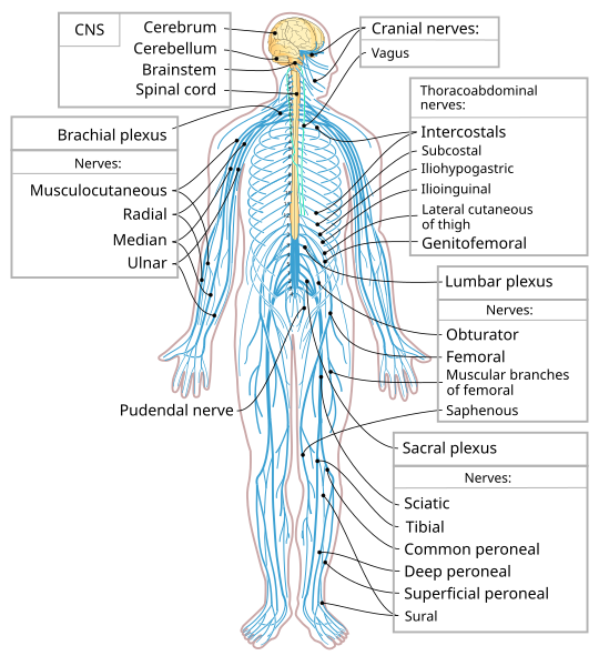
Diagram showing the human central nervous system (brain and spinal cord) contrasted with the peripheral nervous system branching throughout the body. Labels make the boundary between CNS and PNS explicit, reinforcing structural organisation required by the OCR specification. Minimal text keeps the focus on gross anatomy. Source.
Peripheral Nervous System (PNS): The network of sensory and motor neurones linking the CNS with the limbs and organs.
Somatic and Autonomic Nervous Systems
Somatic Nervous System
The somatic system controls voluntary actions, sending impulses to skeletal muscles. It involves one motor neurone directly connecting the CNS to the effector. Reflex arcs such as the knee-jerk reflex also operate through somatic pathways but are involuntary and rapid for protective purposes.
Key features:
Effectors are skeletal muscles.
Usually under conscious control.
Neurotransmitter: acetylcholine.
Autonomic Nervous System (ANS)
The ANS regulates involuntary functions, controlling smooth muscle, cardiac muscle, and glands.
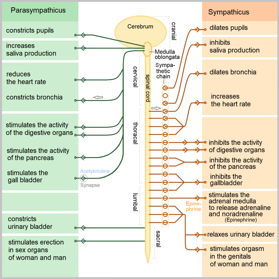
Diagram contrasting sympathetic and parasympathetic outflow and target organs, reinforcing their opposing physiological roles. The layout supports recognition of the ANS as the involuntary division distinct from the somatic system. If additional organ effects are shown, consider them enrichment beyond the OCR core. Source.
It maintains homeostasis by adjusting heart rate, breathing, and digestion automatically.
The ANS has two divisions:
Sympathetic Nervous System (SNS) – prepares the body for fight or flight.
Neurotransmitter: noradrenaline.
Increases heart rate, dilates pupils, reduces digestive activity.
Parasympathetic Nervous System (PNS) – promotes rest and digest functions.
Neurotransmitter: acetylcholine.
Slows heart rate, stimulates digestion, and conserves energy.
Autonomic Nervous System (ANS): The part of the nervous system controlling involuntary actions, divided into sympathetic and parasympathetic divisions.
Comparison of Somatic and Autonomic Systems
Somatic: single motor neurone to effectors.
Autonomic: two neurones connected by a synapse in a ganglion.
Somatic: voluntary skeletal control.
Autonomic: involuntary smooth and cardiac control.
Gross Structure of the Brain
The brain is the most complex organ in the human body, responsible for thought, coordination, homeostasis, and emotion. It is protected by the skull, meninges, and cerebrospinal fluid (CSF), which cushions and nourishes neural tissue.
Main Regions of the Brain
The OCR specification requires understanding of the cerebrum, cerebellum, medulla oblongata, hypothalamus, and pituitary gland.
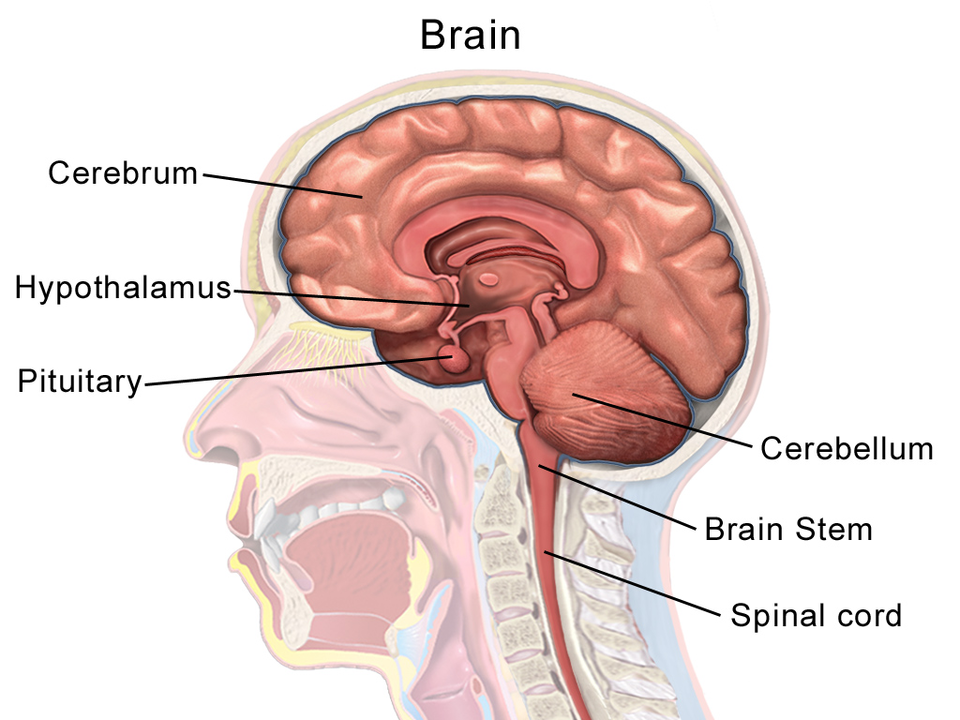
Sagittal brain diagram highlighting the cerebrum (cortical mantle), cerebellum, brainstem/medulla oblongata, hypothalamus, and pituitary. Labels correspond to functions discussed (coordination, autonomic control, and neuroendocrine integration). Extra minor labels for neighbouring structures may appear, but the key OCR-listed regions are prominent. Source.
Cerebrum
The cerebrum is the largest brain region, divided into two cerebral hemispheres, connected by the corpus callosum. Each hemisphere controls the opposite side of the body.
Functions of the cerebrum:
Sensory areas: receive and interpret sensory input (e.g., vision, hearing).
Motor areas: send impulses to effectors for movement.
Association areas: integrate information for learning, reasoning, and emotion.
Cerebrum: The largest part of the brain responsible for sensory perception, voluntary movement, and higher cognitive processes.
The surface of the cerebrum, the cerebral cortex, is highly folded to increase surface area for synaptic connections. Different lobes specialise in particular functions:
Frontal lobe – decision making, reasoning, movement.
Parietal lobe – sensory processing and spatial awareness.
Temporal lobe – hearing, memory, language.
Occipital lobe – vision.
Cerebellum
Located beneath the cerebrum, the cerebellum coordinates movement, balance, and posture. It receives input from the inner ear, muscles, and eyes to ensure smooth, accurate motion.
Functions include:
Maintaining muscle tone and posture.
Coordinating voluntary movements.
Learning motor skills such as playing an instrument or cycling.
Cerebellum: The part of the brain responsible for coordinating balance, posture, and smooth muscular movement.
Medulla Oblongata
The medulla oblongata lies at the base of the brainstem, connecting the brain to the spinal cord. It contains vital control centres for autonomic reflexes essential for survival.
Key regulatory centres:
Cardiac centre – controls heart rate and force of contraction.
Vasomotor centre – adjusts blood vessel diameter and blood pressure.
Respiratory centre – regulates breathing rate and depth.
Medulla Oblongata: The part of the brainstem controlling involuntary processes such as heart rate, breathing, and blood pressure.
Hypothalamus
The hypothalamus is a small but crucial structure below the thalamus, linking the nervous and endocrine systems. It maintains internal balance through homeostatic regulation.
Functions:
Regulates body temperature, hunger, and thirst.
Monitors blood water potential and glucose concentration.
Controls circadian rhythms (sleep–wake cycle).
Coordinates the autonomic nervous system.
Stimulates the pituitary gland to release hormones.
Hypothalamus: A brain region that regulates homeostasis and links the nervous and endocrine systems via control of the pituitary gland.
Pituitary Gland
Located beneath the hypothalamus, the pituitary gland is often termed the “master gland” because it secretes hormones that regulate other endocrine glands.
Structure:
Anterior pituitary – releases hormones such as ACTH, FSH, LH, and growth hormone.
Posterior pituitary – stores and releases ADH and oxytocin produced by the hypothalamus.
Key functions:
Controls growth, reproduction, and water balance.
Integrates neural and hormonal control pathways.
Pituitary Gland: The endocrine gland at the brain’s base that releases hormones controlling other glands and bodily processes.
Integration of Nervous System and Brain Function
The brain and nervous system operate as an integrated unit. The CNS interprets and processes information, while the PNS delivers and executes responses. The autonomic divisions ensure involuntary processes such as heartbeat and digestion continue automatically, maintaining homeostasis.
This intricate coordination allows animals to survive, adapt, and respond effectively to their environment through the seamless communication of neural and hormonal systems.
Practice Questions
Question 1 (2 marks)
State two structural differences between the central nervous system (CNS) and the peripheral nervous system (PNS).
Mark Scheme:
1 mark for each correct difference.
CNS consists of the brain and spinal cord; PNS consists of all nerves outside the CNS.
CNS is enclosed within the skull and vertebral column; PNS is not protected by bone.
CNS mainly processes and integrates information; PNS transmits impulses to and from the CNS.
(Max 2 marks)
Question 2 (5 marks)
Explain how different regions of the brain contribute to the coordination and control of body functions. Include the roles of the cerebrum, cerebellum, medulla oblongata, hypothalamus, and pituitary gland in your answer.
Mark Scheme:
Award 1 mark for each distinct point:
Cerebrum: controls voluntary actions; responsible for reasoning, learning, memory, and conscious thought. (1 mark)
Cerebellum: coordinates movement, balance, and posture; ensures smooth muscular activity. (1 mark)
Medulla oblongata: regulates involuntary activities such as heart rate, breathing, and blood pressure. (1 mark)
Hypothalamus: maintains homeostasis; monitors temperature, water potential, and blood glucose; links nervous and endocrine systems. (1 mark)
Pituitary gland: releases hormones that control other endocrine glands and regulate physiological processes such as growth and reproduction. (1 mark)
(Max 5 marks)
FAQ
The hypothalamus contains thermoreceptor cells that monitor the temperature of the blood flowing through it.
When blood temperature deviates from the set point, the hypothalamus triggers corrective mechanisms.
If temperature rises, it stimulates vasodilation and sweating.
If temperature falls, it initiates shivering and vasoconstriction.
It also receives input from peripheral temperature receptors in the skin, allowing rapid response to environmental temperature changes.
The posterior pituitary stores and releases hormones such as ADH and oxytocin that are produced in the hypothalamus.
These hormones travel down neurosecretory cells, showing a direct neural link.
The anterior pituitary, by contrast, is regulated hormonally by releasing factors from the hypothalamus, stimulating or inhibiting specific hormone secretion.
This dual control demonstrates how nervous signals are translated into hormonal regulation.
The cerebellum does not generate motor commands but fine-tunes those initiated by the motor cortex.
It compares intended movements with sensory feedback from muscles, joints, and balance receptors.
If differences are detected, the cerebellum sends corrective signals to adjust timing, strength, and sequence of muscle contractions.
Damage to the cerebellum causes ataxia, resulting in uncoordinated, jerky movements.
The medulla oblongata contains centres that regulate autonomic reflexes vital for life.
The cardiac centre adjusts heart rate and force of contraction.
The vasomotor centre controls blood vessel diameter, influencing blood pressure.
The respiratory centre sets the rhythm and depth of breathing based on blood CO₂ levels.
These centres operate continuously without conscious control, ensuring constant internal conditions.
The two divisions of the autonomic nervous system act in opposition to maintain homeostasis.
The sympathetic system prepares the body for action: increasing heart rate, dilating pupils, and diverting blood to muscles.
The parasympathetic system restores calm: slowing the heart rate, constricting pupils, and stimulating digestion.
This dynamic balance, known as antagonistic control, ensures that body processes remain stable while allowing quick adaptation to changing conditions.

.png)