
OCR Specification focus:
‘Apply Doppler relation fΔ/f = 2v cosθ / c to determine blood speed in patients.’
Ultrasound Doppler techniques enable clinicians to measure blood flow in real time, using frequency shifts produced by moving red blood cells to provide essential diagnostic information about vascular health.
Understanding Doppler Ultrasound
Doppler ultrasound is a diagnostic method that uses high-frequency longitudinal waves to detect motion within the body. When an ultrasound pulse reflects from moving blood, the frequency of the returned wave differs slightly from that sent by the transducer. This phenomenon arises from the Doppler effect, first introduced here as the change in observed wave frequency due to relative motion between a source and an observer.
Doppler Effect: The change in frequency of a wave when there is relative motion between the source emitting the wave and the observer detecting it.
This slight frequency change is extremely valuable in medical imaging because it gives direct quantitative access to blood flow velocity, enabling assessment of circulatory conditions.
The Doppler Frequency Shift
When a transducer emits ultrasound into a blood vessel, red blood cells act as moving reflectors. The transducer emits at a known frequency, and the reflected signal returns with a different frequency, allowing the scanner’s software to calculate the Doppler frequency shift, denoted fΔ.
EQUATION
—-----------------------------------------------------------------
Doppler Relation (Blood Speed) = fΔ / f = 2v cosθ / c
fΔ = Doppler frequency shift (Hz)
f = transmitted ultrasound frequency (Hz)
v = blood velocity along the ultrasound path (m s⁻¹)
θ = angle between ultrasound beam and direction of blood flow (degrees or radians)
c = speed of ultrasound in tissue, approximately 1540 m s⁻¹
—-----------------------------------------------------------------
The Doppler relation shows that the measured frequency shift depends on both the speed of blood and the orientation of the blood vessel relative to the transducer. This makes angle estimation an essential part of clinical Doppler examinations.
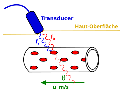
This diagram illustrates Doppler ultrasound of blood flow: an ultrasound transducer sends waves at frequency f₀ into a blood vessel and receives reflections at frequency fᵣ from moving red blood cells. The blood velocity u is shown at an angle θ to the ultrasound beam, highlighting why the cosθ factor appears in the Doppler relation used to determine blood speed. Some labels are in German, but the geometry and physical quantities remain clear and directly support the OCR specification. Source.
Significance of the Doppler Angle
The angle θ appears in the Doppler relation through the cosθ factor, which adjusts how effectively motion contributes to the observed frequency shift. Clinicians aim to keep θ as small as possible because this aligns the ultrasound beam more closely with the direction of flow, increasing accuracy.
A small θ ensures that cosθ is close to 1, so the measured frequency shift is strong and the calculated velocity is reliable. If θ becomes too large, particularly beyond about 60°, cosθ reduces sharply, meaning that even a tiny error in angle measurement causes a large velocity error. This is why the correct transducer orientation is a crucial skill in ultrasound practice.
Components and Operation of Doppler Ultrasound
To make use of Doppler techniques, an ultrasound system employs a specialised piezoelectric transducer capable of both sending and receiving high-frequency sound pulses. These transducers convert electrical pulses into sound and then convert reflected sound into electrical signals for interpretation.
Key operational steps include:
Emission of ultrasound pulses at a precisely controlled frequency.
Interaction with moving blood, where red blood cells reflect the sound.
Receiving the altered waves, containing frequency shifts related to motion.
Processing by onboard signal analysis, which isolates fΔ from the returning signal.
Displaying results via velocity–time traces or colour-coded flow maps.
Between these stages, sophisticated filtering removes unwanted echoes from stationary tissues so that only reflections from moving blood contribute to the Doppler output.
Types of Doppler Presentation
Doppler ultrasound can be presented in several formats that map motion in different ways, enhancing both qualitative and quantitative analysis.
Spectral Doppler
This mode displays a graph of velocity against time. Peaks and troughs in the trace correspond to systolic and diastolic flow, respectively. Spectral Doppler is especially useful for quantifying exact blood velocities and diagnosing stenosis, where abnormal narrowing increases flow speed.
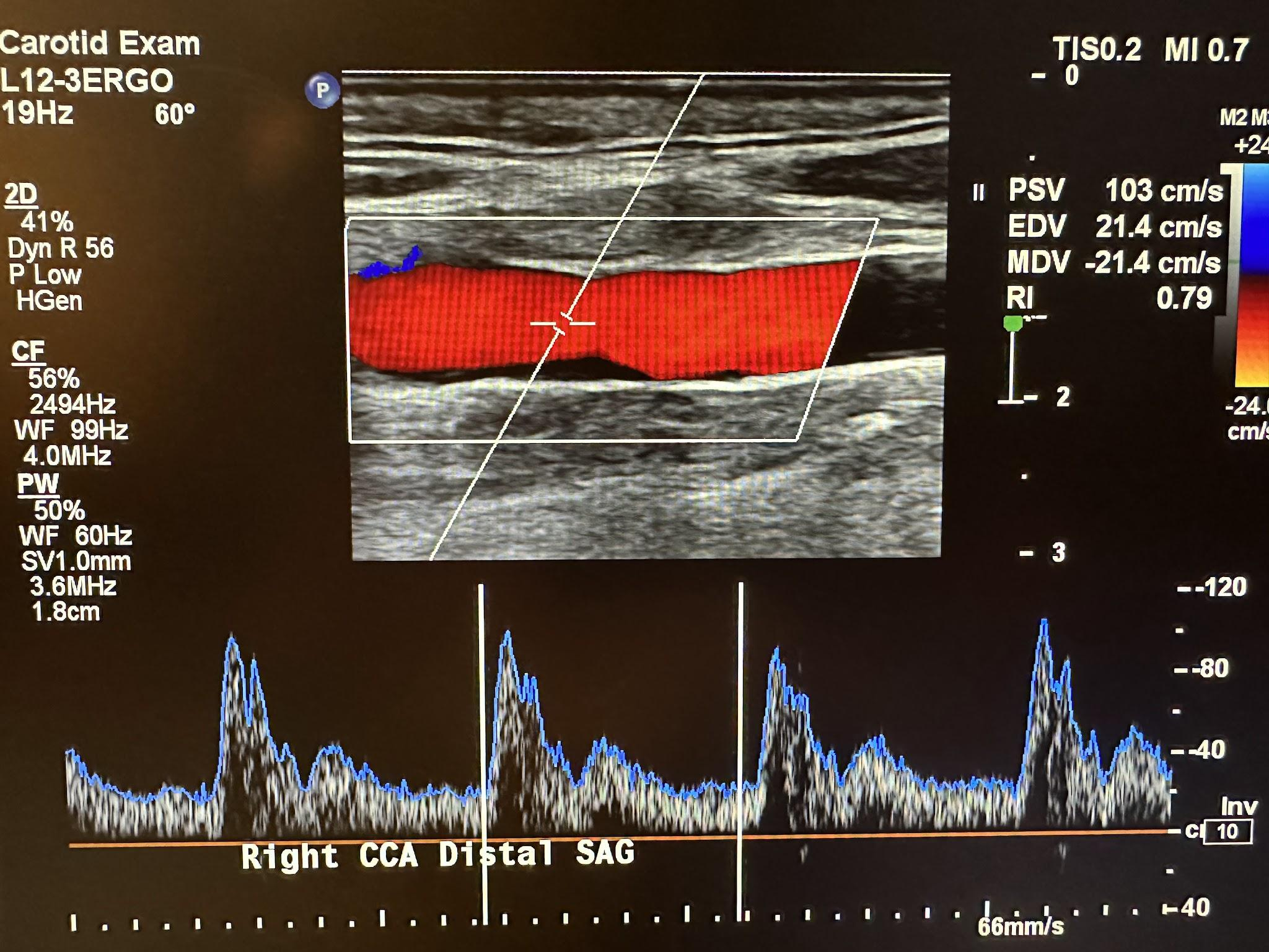
This duplex image shows a longitudinal view of the right common carotid artery with colour Doppler indicating blood flowing through the vessel, and a spectral Doppler trace displaying velocity against time. The labelled systolic and diastolic velocities show how Doppler shifts translate into blood speed measurements using the Doppler relation. Additional machine parameters are visible but extend beyond the OCR syllabus, offering realistic clinical context. Source.
Colour Doppler
Here, the ultrasound system overlays colour on a two-dimensional anatomical image. The colours represent direction and magnitude of flow, commonly with red for flow towards the transducer and blue for flow away. Although less precise than spectral Doppler, it provides rapid visual assessment of vascular patterns.
Power Doppler
This variant shows the strength of the Doppler signal rather than velocity or direction. It is more sensitive to low-speed flows, making it useful in small vessels, though it does not convey directional information.
A normal sentence must appear here before introducing any new definition or equation block, ensuring clear separation in explanation flow.
Red Blood Cell Scatterer: A moving microscopic reflector that returns ultrasound pulses and produces frequency shifts used in Doppler measurements.
Clinical Applications
Doppler ultrasound plays a central role in assessing cardiovascular function because it provides real-time measurement of flow patterns and magnitude. Clinically, it is used to:
Evaluate arterial narrowing (stenosis) by detecting elevated velocities.
Assess venous flow to diagnose thrombosis.
Monitor foetal circulation during pregnancy.
Measure heart valve performance by identifying abnormal regurgitation or turbulence.
Each of these relies fundamentally on accurate interpretation of Doppler frequency shifts through the OCR-required relation fΔ/f = 2v cosθ / c.
Practice Questions
Question 1 (2 marks)
An ultrasound transducer is used to measure blood flow in an artery. The Doppler frequency shift detected is proportional to the component of blood velocity along the ultrasound beam.
Explain why the angle between the beam and the direction of blood flow must be kept as small as possible during Doppler measurements.
Question 1 (2 marks)
1 mark: States that the Doppler frequency shift depends on cos(theta) or the velocity component along the beam.
1 mark: States that small angles maximise cos(theta) (close to 1), giving a larger and more accurate frequency shift / reducing percentage error.
Question 2 (5 marks)
A Doppler ultrasound scanner operates at a transmitted frequency of 3.5 MHz. The ultrasound beam makes an angle of 45 degrees to the direction of blood flow in a patient’s artery.
(a) State the Doppler relation used to determine blood speed from the measured frequency shift. (1 mark)
(b) Explain how the Doppler frequency shift is produced when ultrasound pulses interact with moving red blood cells. (2 marks)
(c) Discuss why inaccurate knowledge of the beam–flow angle can lead to significant errors in calculated blood velocity, and describe how operators minimise such errors in practice. (2 marks)
Question 2 (5 marks)
(a) 1 mark
States the Doppler relation: fΔ / f = 2v cos(theta) / c (symbols may be rearranged or expressed verbally).
(b) 2 marks
1 mark: States that ultrasound waves reflect from moving red blood cells.
1 mark: Explains that motion of the cells causes a change in observed frequency due to the Doppler effect.
(c) 2 marks
1 mark: Explains that errors in estimating theta cause large errors in cos(theta), especially at larger angles, leading to inaccurate velocity calculations.
1 mark: States a practical method to minimise this error, such as aligning the beam as parallel as possible to blood flow, using standard angle settings (e.g., 30–60 degrees), or using built-in angle correction tools.
FAQ
Arteries typically produce larger Doppler shifts because blood moves at higher velocities due to the pulsatile pressure generated by the heart. Veins have lower and more continuous flow, giving smaller shifts.
Other factors also contribute:
Arteries may be positioned so the insonation angle is easier to optimise.
Venous flow is more affected by respiration, causing variability in measured shifts.
Turbulence causes a wide range of red blood cell velocities, resulting in a broadened and noisy spectral waveform.
This leads to:
Loss of a clear velocity envelope
Increased spectral spread
Difficulty identifying peak systolic velocity
Turbulence is common downstream of stenoses and can be an indicator of abnormal flow conditions.
Aliasing occurs when the frequency shift exceeds the system’s Nyquist limit, causing the displayed waveform to appear wrapped or inverted.
It is more likely to occur when:
Blood velocities are high
The sample depth is large
The transmitted frequency is too high
Operators reduce aliasing by lowering frequency, increasing the velocity scale, or switching to continuous-wave Doppler.
Signal strength depends on the number and orientation of red blood cells reflecting ultrasound back to the transducer.
Key influences include:
Haematocrit: fewer red blood cells reduce scattering
Beam alignment: signals are stronger when cells move towards or away from the probe
Attenuation: deeper vessels weaken returning echoes
Vessel size: larger vessels generally provide clearer Doppler signals
Ultrasound gel eliminates air between the probe and skin, preventing reflection losses and ensuring efficient transmission of pulses.
At tissue interfaces, differences in acoustic impedance affect how much energy reaches deeper vessels. Excessive reflection at boundaries can reduce the amplitude of Doppler-shifted echoes, making blood flow harder to detect.
