

Edexcel Syllabus focus:
'Understand how models, such as the fluid mosaic model, interpret data to develop scientific explanations of membrane structure and properties.'
Cell membranes are too small and complex to understand by direct observation alone, so biologists use evidence-based models to explain their structure, behavior, and properties and to refine explanations as new data appears.
Scientific models in biology
A scientific model helps scientists explain structures or processes that are difficult to observe directly. In biology, models are especially useful for microscopic structures such as cell membranes.
Scientific model: A simplified representation of a real structure or process, based on evidence, that is used to explain observations and make predictions.
A model is not a perfect copy of reality. Instead, it selects the most important features needed to explain the available data. As better evidence is collected, the model may be changed or replaced.
For membranes, scientists had to interpret indirect evidence from techniques such as microscopy, chemical analysis, and labeling experiments. This is why membrane structure is understood through a model rather than by simple visual description.
The fluid mosaic model
The fluid mosaic model is the accepted scientific explanation for membrane structure. It describes the membrane as a phospholipid bilayer with proteins associated with it in a dynamic arrangement.
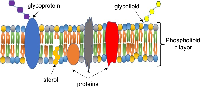
Diagram of the fluid mosaic model showing a phospholipid bilayer with embedded membrane proteins and other components (e.g., sterols and carbohydrate-containing molecules). This visual reinforces how “mosaic” refers to the patchwork distribution of different molecules, while “fluid” reflects lateral movement within the bilayer. Source
Fluid mosaic model: A model of membrane structure in which phospholipids form a bilayer and proteins are distributed within it, with some components able to move sideways through the membrane.
The word fluid shows that parts of the membrane are able to move. Phospholipid molecules can move laterally within the layer, so the membrane is flexible rather than rigid.
The word mosaic shows that the membrane contains a mixture of different components arranged in a patchwork pattern. Proteins are not spread as one continuous sheet. Instead, they are scattered through or attached to the bilayer.
This model explains membrane structure more effectively than earlier ideas because it fits a wider range of evidence. It also explains important membrane properties, including flexibility, selective permeability, and the movement of some membrane components.
How evidence led to the model
Scientific models develop from data. Earlier membrane models suggested that proteins formed continuous layers on the outside of the membrane. These ideas matched some early microscopic observations, but later evidence showed that this explanation was incomplete.
Electron microscopy gave scientists images of membranes with a layered appearance. This supported the idea that membranes had an organized structure, but it did not show exactly how proteins and lipids were arranged.
Later, freeze-fracture microscopy provided stronger evidence. In this method, membranes are split through the middle of the bilayer. The fractured surfaces showed particles embedded within the membrane. These particles were interpreted as proteins, suggesting that proteins are inserted into the bilayer rather than forming separate outer coats.
Other experiments supported the idea of membrane fluidity. When membrane proteins were tagged with fluorescent markers and cells were fused, the markers gradually mixed across the membrane surface. This showed that proteins could move sideways within the membrane.
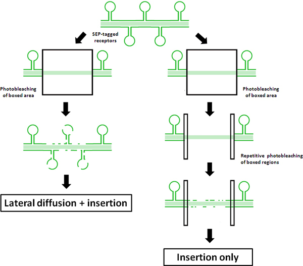
Example fluorescence recovery/photobleaching (FRAP/related) data used to quantify lateral diffusion of membrane-associated proteins in living cells. The recovery of fluorescence over time illustrates that labeled molecules move within the plane of the membrane, providing experimental support for the membrane’s “fluid” behavior. Source
Chemical evidence also supported the model. Membranes were found to contain large amounts of phospholipid, which has hydrophilic and hydrophobic regions. This made the bilayer arrangement a logical explanation, because the hydrophobic parts face inward away from water, while the hydrophilic parts face the watery environments on either side.
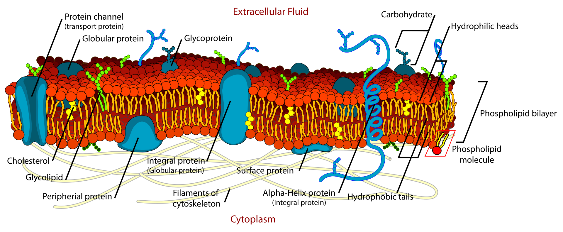
Detailed cross-sectional schematic of the plasma membrane, labeling the hydrophilic phospholipid heads, hydrophobic fatty-acid tails, and major membrane protein types. It supports the evidence-based logic of bilayer formation in water and clarifies how proteins can be embedded within (integral) or associated with (peripheral) the membrane. Source
Together, these results did not simply describe a membrane. They had to be interpreted. The fluid mosaic model was developed as the best explanation that matched the evidence available.
How the model explains membrane properties
Because the membrane is described as fluid, the model explains why membranes are flexible. Cells can change shape, vesicles can form, and membranes can fuse and reseal.
Because the membrane is a bilayer with a hydrophobic interior, the model explains selective permeability. Some substances can pass through easily, while others cannot. Small nonpolar molecules cross more readily than large or charged substances.
Because proteins are part of the mosaic, the model explains why membranes have many different functions. Different membranes can contain different sets of proteins, allowing transport, recognition, or enzymatic activity.
The model also explains why membrane properties are not fixed. The exact arrangement of proteins and lipids can vary between cells and between different membranes in the same organism. This variation changes how the membrane behaves while still fitting the same overall model.
Why the fluid mosaic model is a scientific model
The fluid mosaic model is a clear example of how biology uses models to build explanations from data.
It is evidence-based, because it was developed from experimental observations.
It is simplified, because real membranes are more complex than textbook diagrams.
It is predictive, because it helps scientists explain membrane behavior and make new predictions.
It is changeable, because new evidence can refine the model.
This last point is important. Modern research shows that membranes are not always as uniform as early diagrams suggest. Some proteins are restricted in their movement, and some regions of the membrane may be more organized than others. However, this does not make the model wrong. It shows that scientific models are improved when new evidence becomes available.
The fluid mosaic model remains useful because it captures the main features needed to explain membrane structure and properties, while also allowing for revision as scientific understanding develops.
Practice Questions
Explain what is meant by the terms fluid and mosaic in the fluid mosaic model of membrane structure. (2 marks)
fluid = phospholipids and/or some proteins can move sideways within the membrane (1)
mosaic = proteins are scattered or embedded among the phospholipids in a patchwork arrangement (1)
Explain how the fluid mosaic model acts as a scientific model and how evidence has been used to support or modify it. (5 marks)
a scientific model is a simplified explanation based on evidence / used to explain observations (1)
membrane described as a phospholipid bilayer with proteins associated with it (1)
microscopy evidence supported an organized membrane structure (1)
freeze-fracture evidence showed proteins embedded within the bilayer (1)
movement studies / labeled proteins supported membrane fluidity or new evidence showed the model can be refined, for example some proteins are less mobile than first thought (1)
FAQ
Membrane diagrams are designed to show relationships between parts, not exact size.
If they were drawn to scale:
the phospholipid bilayer would look extremely thin
many proteins would appear much larger than the surrounding lipids
important details would be too small to label clearly
So textbook images are simplified visual models, not exact replicas.
Artificial membranes let scientists control variables more carefully than in living cells.
They can:
change lipid composition
add or remove specific proteins
test how membranes behave under different conditions
This helps scientists decide which features of the membrane are essential to the model and which depend on the cell type.
Fluorescence methods allow scientists to track molecules in living membranes.
For example, a fluorescent tag can be attached to a membrane protein or lipid. Scientists then observe whether the labeled molecules stay in one place or spread over time.
This gives evidence about:
lateral movement
membrane organization
whether different regions behave differently
These methods are valuable because they study membranes in action, not just in fixed images.
It is still taught because it gives the core framework needed to understand membrane structure.
It remains useful because it correctly emphasizes:
a phospholipid bilayer
proteins associated with the bilayer
movement within the membrane
More advanced findings add detail, but they do not remove the basic value of the model. In science, a model can stay useful even when it is later refined.
The same general model applies broadly, but not every membrane has the same composition.
Different membranes can vary in:
protein content
lipid types
degree of fluidity
surface organization
So the fluid mosaic model is a general explanation, not a claim that all membranes are identical. It provides a shared structure while allowing important differences between cell types and organelles.