
OCR Specification focus:
‘Trachea, bronchi, bronchioles and alveoli contain cartilage, ciliated epithelium, goblet cells, smooth muscle and elastic fibres, each contributing to protection, airway patency and efficient gas exchange.’
The mammalian gas exchange system enables efficient oxygen uptake and carbon dioxide removal through specialised airway structures. Each component has adaptations ensuring protection, airflow control, and effective diffusion of respiratory gases.
Structure of the Mammalian Gas Exchange System
Overview of the Respiratory Tract
Air enters through the nasal cavity, passes via the pharynx and larynx into the trachea, which branches into bronchi, then bronchioles, and finally terminates in clusters of alveoli. This branching system creates a vast surface area essential for efficient gas exchange.
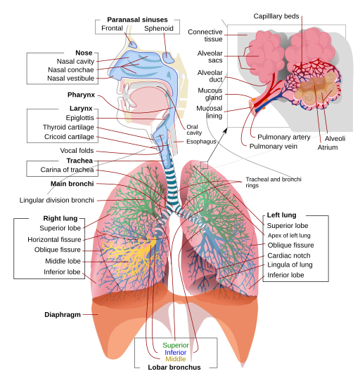
Labelled diagram of the human respiratory system tracing airflow from the trachea to bronchi, bronchioles, and alveoli. The schematic highlights the major conducting airways and the gas-exchange region. Detail is limited to essential labels, supporting the structural overview required by the syllabus. Source.
Trachea
The trachea is a flexible airway supported by C-shaped rings of cartilage.
The cartilage prevents airway collapse during pressure changes of breathing while allowing flexibility for movement of the neck and oesophagus.
The inner lining contains ciliated epithelium and goblet cells, forming the mucociliary escalator, which traps and removes pathogens and dust.
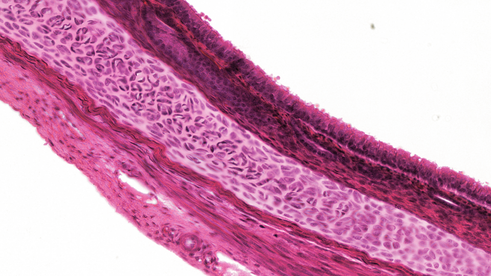
Micrograph of trachea showing the pseudostratified ciliated epithelium at the lumen, submucosal seromucous glands, and a cartilage ring supporting airway patency. The image illustrates the mucociliary surface that protects lower airways. It includes additional tissue layers beyond the syllabus minimum, but these structures help contextualise the epithelium’s protective role. Source.
Ciliated epithelium: A lining of cells bearing microscopic hair-like projections (cilia) that move rhythmically to waft mucus towards the throat for removal.
Bronchi
Each bronchus leads to a lung and has a similar structure to the trachea but with smaller diameter and thinner cartilage plates instead of full rings.
The bronchi continue the defence function of the upper airway using ciliated and mucus-secreting cells.
Smooth muscle present in the walls allows control of airway diameter, adjusting airflow to the lungs.
Bronchioles
The bronchioles are smaller airways lacking cartilage, composed mainly of smooth muscle and elastic fibres.
Constriction or dilation of the smooth muscle regulates airflow distribution.
Elastic fibres permit recoil, maintaining patency and aiding ventilation efficiency.
Terminal bronchioles open into alveolar ducts, connecting to alveolar sacs.
Patency: The state of an airway being open and unobstructed, allowing free passage of air.
Between bronchioles and alveoli, gas exchange efficiency increases due to a dramatic rise in total surface area and a decrease in diffusion distance.
The Alveoli: Site of Gas Exchange
Structure and Adaptations
Alveoli are minute air sacs with an average diameter of about 200–300 μm. Mammalian lungs contain hundreds of millions of alveoli, providing an extensive surface area to volume ratio.
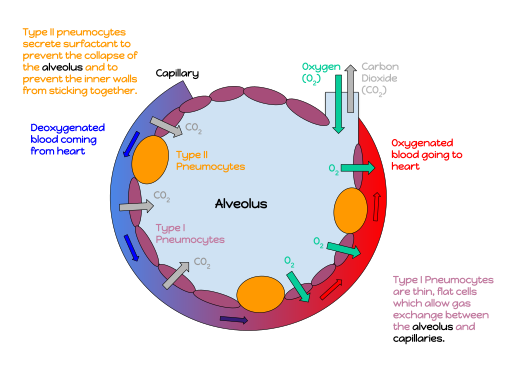
Annotated alveolus showing the single-cell epithelial wall, adjacent capillary network, and the opposing diffusion pathways of oxygen and carbon dioxide. This schematic emphasises large surface area, thin diffusion distance, and maintained gradients. Some versions may also label Type I/II pneumocytes; this is useful context and consistent with the notes, though not strictly required by the specification. Source.
Key structural features include:
Squamous epithelial lining (one cell thick): Minimises diffusion distance.
Elastic fibres: Allow alveoli to stretch during inspiration and recoil during expiration, helping expel air efficiently.
Rich capillary network: Surrounds each alveolus, maintaining a steep concentration gradient for oxygen and carbon dioxide.
Surfactant secretion: Prevents alveolar collapse by reducing surface tension.
Surfactant: A phospholipid-rich fluid secreted by alveolar cells that reduces surface tension, preventing alveolar walls from sticking together during exhalation.
Diffusion Process
Gas exchange across the alveolar membrane occurs by simple diffusion down concentration gradients.
Oxygen diffuses from the alveolar air (high concentration) into capillary blood (low concentration).
Carbon dioxide diffuses in the opposite direction, from blood to alveolar air.
Efficient exchange depends on:A large surface area of alveoli.
Thin exchange surfaces (around 0.5 µm total thickness).
A continuous blood flow maintaining concentration gradients.
Ventilation movements refreshing alveolar air.
Cellular Components of the Gas Exchange Surfaces
Epithelial Cells
Two main types of epithelial cells are present in alveoli:
Type I pneumocytes: Extremely thin, forming most of the alveolar wall for diffusion.
Type II pneumocytes: Secrete surfactant and can divide to replace damaged cells.
Capillary Endothelium
The endothelial cells of pulmonary capillaries are also thin and permeable, allowing gases to diffuse rapidly between air and blood. Blood flow through capillaries is slow, increasing exposure time for diffusion.
Supporting Tissues
Elastic and collagen fibres provide structural integrity. Elastic fibres, in particular, are vital for elastic recoil, a passive force driving exhalation.
Elastic recoil: The tendency of stretched elastic fibres in the lung tissue to return to their original shape, aiding expulsion of air during exhalation.
Functional Integration of Airway Components
Role of Cartilage
Cartilage ensures airway stability, preventing collapse under changing pressure conditions. Its discontinuous C-shape in the trachea allows flexibility and permits oesophageal expansion during swallowing.
Role of Smooth Muscle
Smooth muscle adjusts airflow resistance. Contraction narrows the airway (bronchoconstriction), reducing airflow — important in reflex responses or allergic reactions. Relaxation widens airways (bronchodilation) to maximise airflow during exercise.
Role of Ciliated Epithelium and Goblet Cells
Together they provide a protective mucociliary barrier:
Goblet cells produce mucus to trap particles.
Cilia move mucus upwards toward the throat for swallowing.
This helps maintain clean, pathogen-free airways, safeguarding delicate alveoli.
Role of Elastic Fibres
Elastic fibres allow stretch and recoil of airways and alveoli, crucial for maintaining pressure changes that facilitate ventilation.
Importance for Efficient Gas Exchange
An efficient mammalian gas exchange system balances structural protection with functional optimisation:
Cartilage maintains open airways.
Smooth muscle regulates airflow.
Ciliated epithelium and goblet cells defend against pathogens.
Elastic fibres and extensive capillary networks enable effective diffusion.
Together, these features ensure that oxygen uptake and carbon dioxide removal occur rapidly and continuously to meet the high metabolic demands of mammals.
Practice Questions
Question 1 (2 marks)
Describe two structural features of the trachea that help to maintain efficient airflow in the mammalian gas exchange system.
Mark scheme:
C-shaped rings of cartilage prevent the trachea from collapsing while allowing flexibility (1 mark).
Ciliated epithelium and goblet cells form the mucociliary escalator that traps and removes dust and microorganisms, keeping airways clear (1 mark).
(2 marks total)
Question 2 (5 marks)
Explain how the structure of the alveoli and their surrounding capillaries ensures efficient gas exchange in mammals.
Mark scheme:
Large number of alveoli provide a large surface area for diffusion (1 mark).
Alveolar walls are one cell thick (squamous epithelium) giving a short diffusion distance (1 mark).
Capillary walls are also one cell thick allowing rapid diffusion between air and blood (1 mark).
Extensive capillary network maintains a steep concentration gradient by constant blood flow (1 mark).
Elastic fibres in alveolar walls allow expansion during inspiration and recoil during expiration to maintain ventilation and concentration gradients (1 mark).
(5 marks total)
FAQ
During exercise, smooth muscle relaxes under the influence of adrenaline, causing bronchodilation. This widens the airways, reduces resistance, and increases airflow to meet higher oxygen demands.
In contrast, during allergic reactions or asthma, smooth muscle contracts in response to histamine or other inflammatory mediators, leading to bronchoconstriction. This narrows the airways, restricts airflow, and can cause wheezing or shortness of breath.
Newborns, particularly premature infants, may lack sufficient pulmonary surfactant, a phospholipid-rich fluid that reduces surface tension in the alveoli.
Without it, alveoli tend to collapse (atelectasis) after exhalation, making reinflation difficult. This leads to infant respiratory distress syndrome (IRDS).
Administering artificial surfactant and mechanical ventilation helps stabilise alveoli and ensures efficient gas exchange in neonatal care.
Alveolar macrophages are immune cells that patrol the alveolar surface, providing defence against inhaled pathogens and particles.
They engulf and digest debris or microorganisms through phagocytosis.
They help maintain sterile conditions without disrupting the delicate gas exchange surfaces.
After performing their function, they migrate towards the bronchioles, where they are swept up by the mucociliary escalator and expelled or swallowed.
This mechanism complements structural defences, maintaining healthy alveoli.
Elastic fibres not only assist in passive exhalation by recoil but also play a role in even air distribution.
During inhalation, they stretch to accommodate expanding alveoli, preventing overinflation of certain regions.
As they recoil, they help push air out uniformly, preventing trapped air pockets.
Loss of elasticity, as in emphysema, reduces this efficiency, causing difficulty in exhalation and poor gas exchange.
The gradient for oxygen and carbon dioxide diffusion is sustained by two main processes:
Ventilation constantly replenishes alveolar air, keeping oxygen concentration high and carbon dioxide low.
Continuous blood flow through pulmonary capillaries removes oxygenated blood and brings deoxygenated blood with high carbon dioxide content.
Together, these processes ensure diffusion remains efficient throughout each breathing cycle, even during increased metabolic activity.

_histology_cross-section.png)