
OCR Specification focus:
‘Dissect and draw bony fish gills or insect tracheae, and examine histology slides to identify the tissues and adaptations of exchange surfaces.’
Understanding and observing exchange surfaces through practical dissections and microscopy enhances comprehension of their structural adaptations for efficient gas exchange in animals and provides essential biological skills.
Dissections of Exchange Surfaces
Importance of Dissections
Dissections provide direct observation of the structure-function relationship in respiratory systems, reinforcing theoretical understanding of gas exchange mechanisms in animals. Ethical practice and precise technique are essential.
Safety and Ethical Considerations
Before any dissection:
Ensure proper personal protective equipment (PPE) such as gloves, goggles, and lab coats.
Use sterile instruments and dispose of specimens safely following biological waste protocols.
Follow ethical sourcing: all specimens must be ethically obtained and treated humanely before death.
Avoid unnecessary tissue damage to preserve structural clarity for educational observation.
Dissecting Bony Fish Gills
Bony fish, such as trout, provide excellent examples of efficient aquatic gas exchange systems. The gills demonstrate adaptations that maintain a steep diffusion gradient through countercurrent flow of water and blood.
Procedure Overview:
Place the fish on a dissecting tray with its operculum facing upwards.
Carefully lift and remove the operculum to expose the gill arches.
Observe gill filaments extending from each arch, covered in secondary lamellae.
Identify afferent and efferent blood vessels, noting their proximity for effective diffusion.
Draw a labelled biological diagram, showing cartilage support, filament arrangement, and vascularisation.
Carefully lift the operculum to reveal the gill arches, filaments and lamellae.
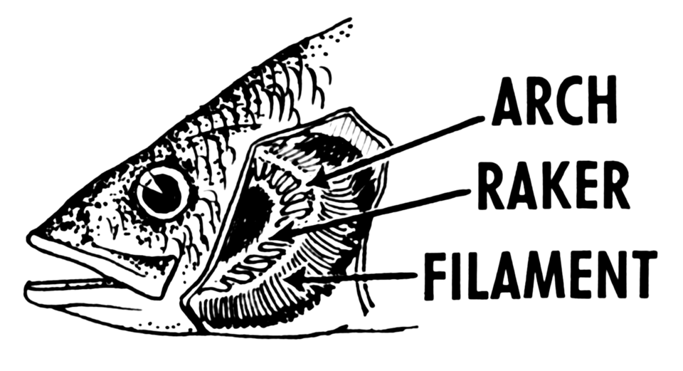
Labeled line drawing of a bony fish gill showing the gill arch supporting multiple filaments that bear closely spaced lamellae. This arrangement maximises surface area and minimises diffusion distance, matching the functional adaptations discussed in the dissection protocol. Suitable as a template for labelled student drawings. Source.
Key Observations in Fish Gills
Large surface area provided by lamellae enhances gas diffusion.
Thin epithelial layers minimise diffusion distance.
Countercurrent flow maintains a steep oxygen concentration gradient, maximising oxygen uptake.
Rich blood supply within lamellae facilitates efficient gas transport.
Countercurrent Flow: The opposite movement of water and blood across gill lamellae that maintains a continuous diffusion gradient for oxygen uptake.
Fish dissections illustrate how structure supports physiological efficiency, a fundamental concept in comparative respiratory biology.
Dissection of Insect Tracheal Systems
Overview of Insect Gas Exchange
Insects exchange gases through a tracheal system, a network of tracheae and tracheoles delivering oxygen directly to tissues without the involvement of blood.
Procedure Overview
Select a large insect (e.g., locust) for visibility of tracheal structures.
Immerse in a dissecting fluid such as saline to prevent desiccation.
Pin out the specimen dorsal side up and remove the exoskeleton gently.
Under magnification, identify white or silvery tracheal tubes branching throughout the body.
Note spiracles, small openings on the thorax or abdomen, which regulate air entry.
Create a labelled drawing of tracheae and tracheoles, indicating their proximity to muscles.
Insects possess spiracles leading into tracheae that branch into tracheoles within tissues, with thoracic and abdominal movements aiding ventilation.
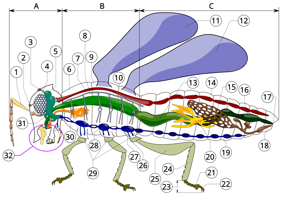
Whole-body diagram of an insect with tracheal tubes (with spiracle) labelled, illustrating entry points and the branching air-tube network. Use it to orient students before zooming in on tracheoles and tracheal fluid in the text. Extra detail (digestive and nervous systems) is present but can be ignored for this subsubtopic. Source.
Key Observations in Insect Tracheae
Spiracles open and close to minimise water loss while enabling gas exchange.
Tracheae are strengthened with chitin rings to prevent collapse.
Tracheoles reach individual cells, containing tracheal fluid that regulates diffusion distance.
Body movements of thorax and abdomen aid ventilation by altering body volume.
Tracheal Fluid: Liquid found at the end of tracheoles that controls the diffusion pathway length for oxygen between air and cells.
These structures exemplify adaptations to terrestrial environments, balancing gas exchange with water conservation.
Microscopy of Exchange Surfaces
Preparing Slides and Observations
Microscopy allows students to identify tissues and observe histological adaptations of gas exchange surfaces. Slides may be prepared in class or examined pre-made for clarity.
Steps for Microscopy:
Use low-power objective to locate the exchange surface.
Switch to high power for detailed examination of tissues such as epithelium, cartilage, smooth muscle, and elastic fibres.
Sketch accurately, noting scale, magnification, and tissue arrangement.
Under the microscope, identify pseudostratified ciliated epithelium with goblet cells, smooth muscle and elastic fibres, and distinguish the type I and type II pneumocytes in alveoli.
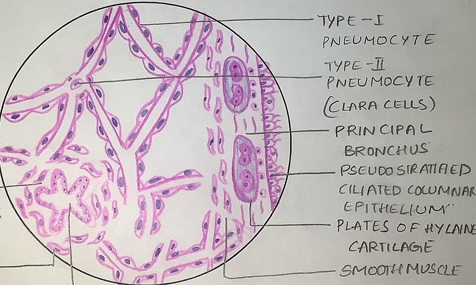
Annotated lung histology showing the respiratory epithelium and the alveolar wall with type I (thin) and type II (surfactant-secreting) pneumocytes. These features underpin thin diffusion distance and surface tension reduction essential for gas exchange. Some panels include contextual labels beyond the minimum syllabus list (e.g., additional alveolar detail). Source.
Tissues in Mammalian Gas Exchange
Microscopy of mammalian lungs (often used as comparison) highlights key features of alveoli, even if not part of this dissection’s focus.
Key tissues visible:
Ciliated epithelium: Moves mucus and trapped particles out of airways.
Goblet cells: Produce mucus for trapping dust and microbes.
Smooth muscle: Controls airway diameter for ventilation.
Elastic fibres: Allow alveoli and airways to stretch and recoil.
Cartilage: Maintains airway patency in trachea and bronchi.
Histology: The microscopic study of tissue structure and organisation to understand functional adaptations.
Microscopy provides direct visual evidence of how tissue types combine to optimise gas exchange efficiency and protection.
Drawing and Recording Biological Observations
Biological Drawing Conventions
Accurate biological drawings are crucial for demonstrating understanding and skill:
Use sharp pencils, clear outlines, and no shading.
Include labels and annotations identifying structures and their functions.
Indicate magnification and scale bars when applicable.
Keep drawings neat and proportionate to observations.
Recording Data and Observations
Record qualitative and quantitative data systematically:
Use observation tables to note structures, functions, and tissue types.
Compare different organisms’ exchange surfaces, focusing on structural adaptations to their environments.
Use scientific terminology consistently to communicate findings accurately.
Skills Developed in Practical Study of Exchange Surfaces
Engaging in dissections and microscopy fosters key biological and analytical skills:
Observation and precision in identifying structures.
Interpretation of how structure relates to function.
Application of ethical practice and biological safety.
Microscopical technique including focusing, slide preparation, and accurate measurement.
Data presentation and drawing accuracy, essential for practical assessments.
These skills form a foundation for higher-level biological research and practical competence, directly aligning with OCR’s emphasis on understanding adaptations and efficiency of exchange surfaces in different organisms.
Practice Questions
Question 1 (2 marks)
During a dissection of a bony fish, a student observes numerous thin lamellae attached to each gill filament.
Explain how this structure aids efficient gas exchange in fish.
Mark Scheme
1 mark: Large surface area provided by many lamellae increases the rate of diffusion.
1 mark: Short diffusion distance due to thin epithelial layers allows rapid gas exchange between water and blood.
Question 2 (5 marks)
Describe how microscopy and dissection can be used to study the structure and adaptations of gas exchange surfaces in animals.
In your answer, include both techniques and the key observations that relate structure to function.
Mark Scheme
Award 1 mark per distinct point up to a maximum of 5 marks:
Use of dissection to expose and observe structures such as fish gills or insect tracheae.
Observation of adaptations: large surface area (lamellae or tracheoles), thin diffusion pathway, and good ventilation/blood flow.
Accurate biological drawing of observed structures with correct labelling and scale.
Use of light microscopy or histology slides to identify tissues such as ciliated epithelium, goblet cells, smooth muscle, and cartilage.
Linking structure to function, e.g. elastic fibres allowing recoil or cilia removing debris to maintain efficiency of gas exchange.
FAQ
Cartilage appears as large, purple-stained, semi-transparent structures with chondrocytes (cartilage cells) in small cavities called lacunae. It forms C-shaped rings in tracheal sections.
Smooth muscle, on the other hand, forms thin bands or layers of elongated spindle-shaped cells with central nuclei. It lacks the clear structural gaps seen in cartilage and may appear between connective tissue and epithelial layers.
Recognising these differences helps students correctly identify the tissues that maintain airway structure and control airway diameter.
Fresh or properly preserved specimens retain the integrity of soft tissues, allowing clearer observation of delicate structures like lamellae and tracheoles.
If tissues dry out or decay, thin membranes collapse, and colour changes obscure the identification of key features such as blood vessels or epithelial linings.
High-quality specimens ensure that students can accurately interpret structural adaptations and make biologically realistic drawings.
Adding shading or colour, which is not allowed in biological drawings.
Using inaccurate proportions—for example, exaggerating the thickness of epithelial layers.
Failing to include labels or scale bars.
Overcrowding the drawing with unnecessary detail instead of focusing on clarity.
Drawings should have clear lines, correct labelling, and reflect the true appearance of the observed specimen for credit in practical assessments.
Accuracy depends on careful sectioning, staining, and mounting:
Use thin sections to allow light penetration and sharp focus under the microscope.
Apply appropriate stains such as haematoxylin and eosin to differentiate cell types and structures.
Remove excess stain and air bubbles before sealing the cover slip.
Observing these steps ensures clear visualisation of epithelial cells, elastic fibres, and capillaries, allowing correct identification during microscopy tasks.
Dissections involve handling real animal tissues, so respecting animal welfare and ethical sourcing is essential.
Specimens must come from animals that were humanely treated and ethically obtained, avoiding unnecessary harm.
Following proper ethical guidelines not only promotes responsible science but also ensures compliance with OCR practical standards, encouraging respect for living organisms and their biological complexity.

.png)