
OCR Specification focus:
‘Know the liver’s gross structure and histology and examine stained sections to identify hepatic tissues and blood vessels.’
The liver is a large, vital organ located in the upper right abdomen that performs multiple metabolic and homeostatic functions, including detoxification, storage, and synthesis. Understanding its structure and histology reveals how these processes are coordinated at both macroscopic and microscopic levels.
Liver Gross Structure
The liver is the largest internal organ in the human body, weighing approximately 1.5 kg in adults. It is divided into two main lobes—the right and left lobes—separated by the falciform ligament, a fold of peritoneum that attaches the liver to the anterior abdominal wall and diaphragm.
Blood Supply
The liver has a unique dual blood supply, receiving both oxygenated and deoxygenated blood:
Hepatic artery – delivers oxygenated blood from the aorta (via the coeliac artery).
Hepatic portal vein – delivers nutrient-rich, deoxygenated blood from the digestive system.
Blood leaves the liver through the hepatic vein, which drains into the inferior vena cava.
This arrangement allows the liver to process substances absorbed from the gut before they reach general circulation.
Microscopic Structure: The Lobule
At the microscopic level, the liver is organised into structural and functional units known as lobules. Each lobule is roughly hexagonal and centred around a central vein.
Components of a Liver Lobule
Hepatocytes – the main liver cells, arranged in plates radiating from the central vein.
Sinusoids – specialised capillaries between hepatocyte plates through which mixed blood (from the hepatic artery and portal vein) flows.
Portal triads – located at each corner of the lobule; each consists of:
A branch of the hepatic artery
A branch of the hepatic portal vein
A bile ductule (part of the bile transport system)
Blood flows from the portal triad towards the central vein, allowing close contact between blood and hepatocytes for metabolic exchange.
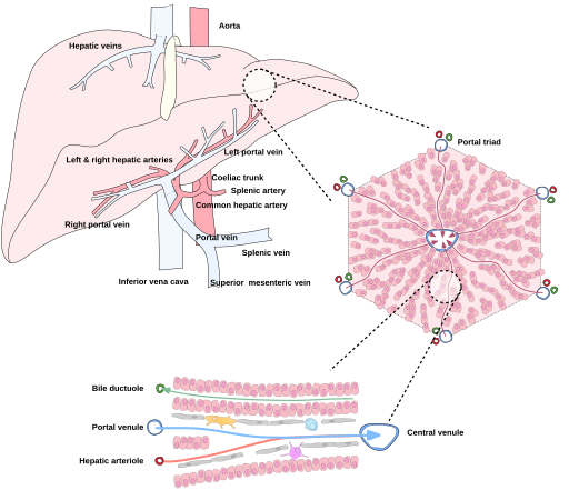
Labeled schematic of a hepatic lobule showing blood entering via branches of the hepatic artery and portal vein at the portal triad and draining to the central vein. The diagram also situates the lobule within the whole liver, aiding orientation to gross anatomy. This directly supports identification of the portal tract and central venule discussed in histology. Source.
Hepatocytes: Structure and Function
Hepatocytes are polygonal epithelial cells that make up approximately 80% of the liver’s mass. They possess:
A large nucleus (sometimes binucleate)
Abundant smooth and rough endoplasmic reticulum
Numerous mitochondria for energy-demanding processes
Microvilli projecting into the space of Disse for increased surface area in exchange
These features enable hepatocytes to perform diverse functions, including protein synthesis, detoxification, bile production, and metabolic regulation.
Hepatocyte: The main functional cell of the liver, responsible for metabolism, detoxification, and synthesis of plasma proteins and bile.
The Sinusoids and Blood Flow
Sinusoids are wide, thin-walled capillaries lined with fenestrated endothelial cells that allow plasma (but not cells) to pass between blood and hepatocytes. Blood from both the hepatic artery and portal vein mixes here before flowing towards the central vein.
Between the endothelium and hepatocytes lies the space of Disse, which facilitates exchange of nutrients, oxygen, and waste products between blood plasma and liver cells.
Specialised Cells of the Sinusoids
Kupffer cells – phagocytic macrophages that line the sinusoids and engulf bacteria, old red blood cells, and debris.
Endothelial cells – regulate permeability and maintain contact with circulating blood.
Stellate (Ito) cells – store vitamin A and can produce collagen in response to injury, contributing to fibrosis in chronic liver disease.
Kupffer cell: A specialised macrophage within liver sinusoids responsible for phagocytosis of pathogens and cellular debris.
The Portal Triad and Bile Canaliculi
Each portal triad represents a junction where blood inflow and bile outflow occur side by side.
Key Features
Hepatic artery branch brings oxygenated blood.
Portal vein branch brings nutrient-rich, deoxygenated blood.
Bile ductule collects bile secreted by hepatocytes.
Between adjacent hepatocytes lie tiny bile canaliculi, forming channels that carry bile away from the centre of the lobule toward the bile ducts in the portal triad.
Bile canaliculus: A microscopic channel formed between hepatocytes that collects bile and transports it toward the bile ductules.
At the lobule periphery, locate a portal triad—a portal venule (PV), hepatic arteriole (HA) and bile ductule (BD)—embedded within connective tissue.
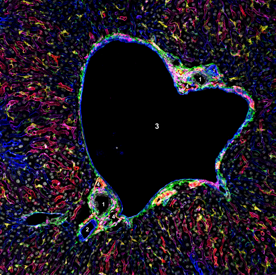
Labelled portal triad micrograph identifying the bile duct (1), hepatic artery (2), portal vein (3) and lymphatic vessels (4). This reinforces recognition of vessel wall thickness differences (artery vs vein) and duct epithelium in situ. The image is from mouse liver, but the microanatomy is directly applicable to human histology at OCR level. Source.
Blood and Bile Flow Directions
Blood and bile flow in opposite directions within a lobule:
Blood: flows from portal triad → sinusoids → central vein.
Bile: flows from hepatocytes → canaliculi → bile ductule.
This counter-current arrangement ensures that hepatocytes along the sinusoids have optimal exposure to both incoming nutrients and outgoing waste products.
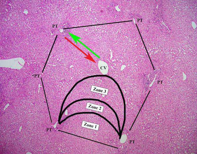
Figure 4 illustrates the classic hepatic lobule with portal tracts at the periphery and a central vein, plus opposing directions of blood and bile flow. Figure 6 shows an H&E micrograph where plates of hepatocytes radiate towards the central vein. Together, they train students to map the schematic onto real tissue. Source.
Histology of the Liver
Under a microscope, a stained section of liver tissue shows:
Central vein in the centre of each lobule.
Radiating cords or plates of hepatocytes.
Sinusoidal spaces between hepatocyte cords.
Portal triads at the periphery with distinct vascular and duct structures.
Histological stains such as haematoxylin and eosin (H&E) are commonly used:
Haematoxylin stains nuclei blue-purple.
Eosin stains cytoplasm pink-red.
This contrast highlights the liver’s architecture and allows identification of cellular and vascular features required in OCR microscopy work.
Adaptations of the Liver for Function
The liver’s histological structure is highly adapted for its metabolic, synthetic, and detoxifying roles:
Extensive blood supply ensures rapid delivery and removal of substances.
Large surface area of hepatocytes facilitates efficient diffusion.
Close contact between hepatocytes and blood plasma via sinusoids and spaces of Disse supports exchange.
Presence of Kupffer cells aids in immune defence.
Bile canaliculi system ensures efficient removal of bile for digestion.
Clinical Relevance of Histological Features
Microscopic examination of liver tissue can reveal disease processes:
Fatty liver – accumulation of lipid droplets within hepatocytes.
Cirrhosis – fibrosis and nodular regeneration disrupting lobule structure.
Hepatitis – inflammation causing cellular damage and infiltration by immune cells.
Such pathological changes are identifiable in stained liver sections, making histology crucial for medical diagnosis and understanding functional impairment.
Summary of Key Structural Relationships
The liver’s gross structure enables blood inflow and outflow via hepatic vessels.
The lobule forms the basic microscopic unit, with hepatocytes performing essential metabolic tasks.
Sinusoids and Kupffer cells mediate filtration and immune protection.
Portal triads coordinate vascular inflow with bile outflow, central to maintaining efficient homeostatic control.
Practice Questions
Question 1 (2 marks)
Name two blood vessels that supply the liver and describe the type of blood each vessel carries.
Mark Scheme:
1 mark for correctly naming each vessel:
Hepatic artery
Hepatic portal vein
1 additional mark for describing the type of blood each carries:
Hepatic artery carries oxygenated blood from the aorta.
Hepatic portal vein carries deoxygenated, nutrient-rich blood from the digestive system.
(Max 2 marks)
Question 2 (5 marks)
Describe and explain how the structure of a liver lobule is adapted to its function in the body.
Mark Scheme:
Award marks for any five of the following, with a maximum of one mark per valid point:
Hexagonal structure of the lobule allows efficient organisation of cells around a central vein for blood flow.
Plates of hepatocytes radiate from the central vein to increase surface area for exchange with blood.
Sinusoids between hepatocyte plates allow close contact between blood and liver cells for metabolic exchange.
Fenestrated endothelium of sinusoids permits diffusion of plasma components between blood and hepatocytes.
Kupffer cells in sinusoids remove bacteria and debris, maintaining blood quality.
Portal triads at lobule corners supply blood (from hepatic artery and portal vein) and remove bile via bile ductules.
Bile canaliculi between hepatocytes efficiently transport bile away from cells towards bile ducts.
Overall structure ensures efficient detoxification, metabolism, and bile secretion by maximising contact between blood and liver tissue.
FAQ
The hepatic lobule is the traditional structural unit of the liver, centred on the central vein with portal triads at each corner. It shows the direction of blood flow from the periphery to the centre.
The hepatic acinus, however, is a more functional unit, centred around a portal triad, describing how oxygen and nutrients are distributed to hepatocytes. It divides the tissue into zones 1–3, from most oxygenated (near the portal triad) to least (near the central vein).
Lobule zones differ in exposure to oxygen and nutrients:
Zone 1 (periportal): Closest to the portal triad; receives the most oxygen and nutrients. Active in gluconeogenesis, urea formation, and oxidation reactions.
Zone 3 (centrilobular): Near the central vein; lowest oxygen levels. Specialises in glycolysis and detoxification.
Zone 3 hepatocytes are more vulnerable to hypoxia and toxic damage, as they have limited oxygen but higher exposure to metabolic by-products.
Hepatic sinusoids differ structurally and functionally:
They are wider and irregularly shaped compared to normal capillaries.
Their endothelial lining is fenestrated, allowing plasma but not blood cells to pass.
They lack a complete basement membrane, facilitating exchange between blood and hepatocytes through the space of Disse.
Kupffer cells are attached to the sinusoid walls, providing immune surveillance and debris removal.
These adaptations make sinusoids ideal for high-volume metabolic exchange.
Hepatocytes have a remarkable ability to undergo mitosis to replace damaged cells. Following injury or partial hepatectomy:
Growth factors such as hepatocyte growth factor (HGF) and epidermal growth factor (EGF) stimulate division.
Kupffer cells release cytokines like interleukin-6 to activate hepatocyte proliferation.
The liver can restore its original mass within weeks if the underlying structure remains intact.
However, chronic damage may lead to fibrosis and cirrhosis, impairing regeneration.
The space of Disse lies between the sinusoidal endothelium and hepatocytes. It allows exchange of plasma components with liver cells for metabolism.
Functions include:
Enabling diffusion of nutrients, hormones, and waste between blood and hepatocytes.
Providing location for microvilli of hepatocytes, increasing surface area for exchange.
Housing stellate (Ito) cells, which store vitamin A and produce extracellular matrix components.
Disruption of this space, such as by collagen deposition in fibrosis, severely reduces liver efficiency.
