
OCR Specification focus:
‘Osmoreceptors in the hypothalamus, posterior pituitary release of ADH, and ADH action on collecting duct walls regulate blood water potential.’
The control of blood water potential is vital for maintaining homeostasis, ensuring cells neither shrink nor swell excessively. This process is regulated hormonally by antidiuretic hormone (ADH).
Regulation of Blood Water Potential
The blood water potential determines the balance between water and solutes in plasma. It must remain within a narrow range to maintain cellular function and osmotic stability. When this balance changes, osmoregulation—the control of water content and solute concentration—ensures that the internal environment remains constant despite fluctuations in external conditions.
Osmoregulation: The maintenance of a constant internal water potential in the blood and tissue fluids, despite changes in external water or solute levels.
Changes in blood water potential may arise from factors such as dehydration, excessive sweating, high salt intake, or overhydration. These variations are detected by osmoreceptors, triggering responses that adjust water reabsorption in the kidneys.
The Role of Osmoreceptors in the Hypothalamus
Osmoreceptors are specialised sensory neurones located in the hypothalamus, a region of the brain that coordinates autonomic and endocrine responses. They are extremely sensitive to small changes in blood water potential.
When blood water potential decreases (becomes more negative), due to water loss or increased solute concentration, osmoreceptors shrink as water leaves their cytoplasm by osmosis.
When blood water potential increases (becomes less negative), due to water intake or low solute concentration, osmoreceptors swell as water enters by osmosis.
The shrinking or swelling of osmoreceptors influences the activity of neurosecretory cells in the hypothalamus, which control the release of antidiuretic hormone (ADH) from the posterior pituitary gland.
The Posterior Pituitary and ADH Release
The posterior pituitary gland does not synthesise hormones itself; instead, it stores and releases ADH produced in the hypothalamus.
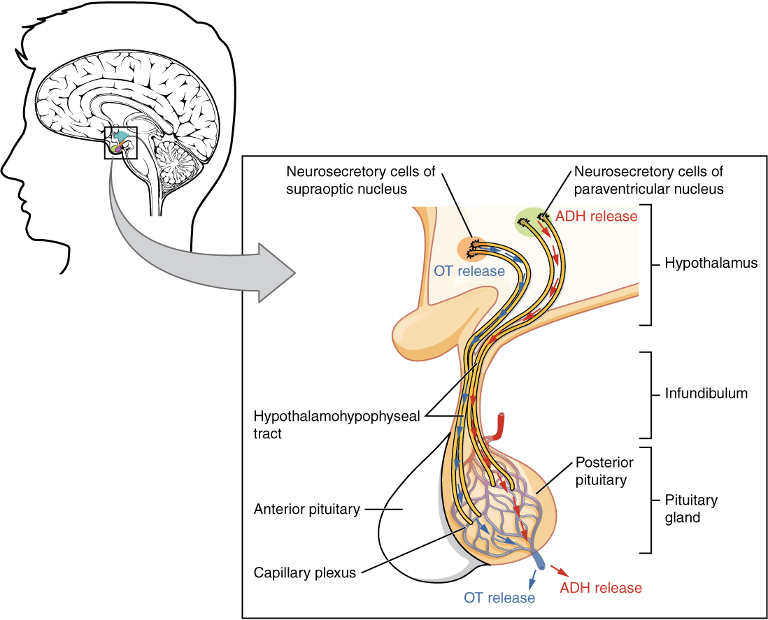
Schematic of the posterior pituitary showing neurosecretory cells in the supraoptic and paraventricular nuclei sending axons through the infundibulum to terminals in the posterior lobe. ADH produced in the hypothalamus is stored and released into the capillary plexus here. Clean labelling emphasises the anatomical route essential for osmoregulatory control. Source.
ADH is synthesised in the supraoptic and paraventricular nuclei of the hypothalamus and transported down axons to the posterior pituitary via neurosecretory vesicles.
When osmoreceptors detect a low blood water potential, they stimulate these neurosecretory cells to release ADH into the bloodstream through exocytosis. The amount of ADH released is proportional to the degree of water imbalance.
In dehydration: High levels of ADH are released to conserve water.
In overhydration: ADH secretion is inhibited to promote water excretion.
Antidiuretic Hormone (ADH): A peptide hormone that increases the permeability of the kidney collecting ducts to water, allowing more water to be reabsorbed into the bloodstream.
Once secreted, ADH travels in the blood to its target organ—the kidney, specifically the distal convoluted tubule and collecting duct.
ADH Action on the Collecting Duct Walls
The main site of ADH action is the collecting duct of the nephron, where water reabsorption is finely controlled.
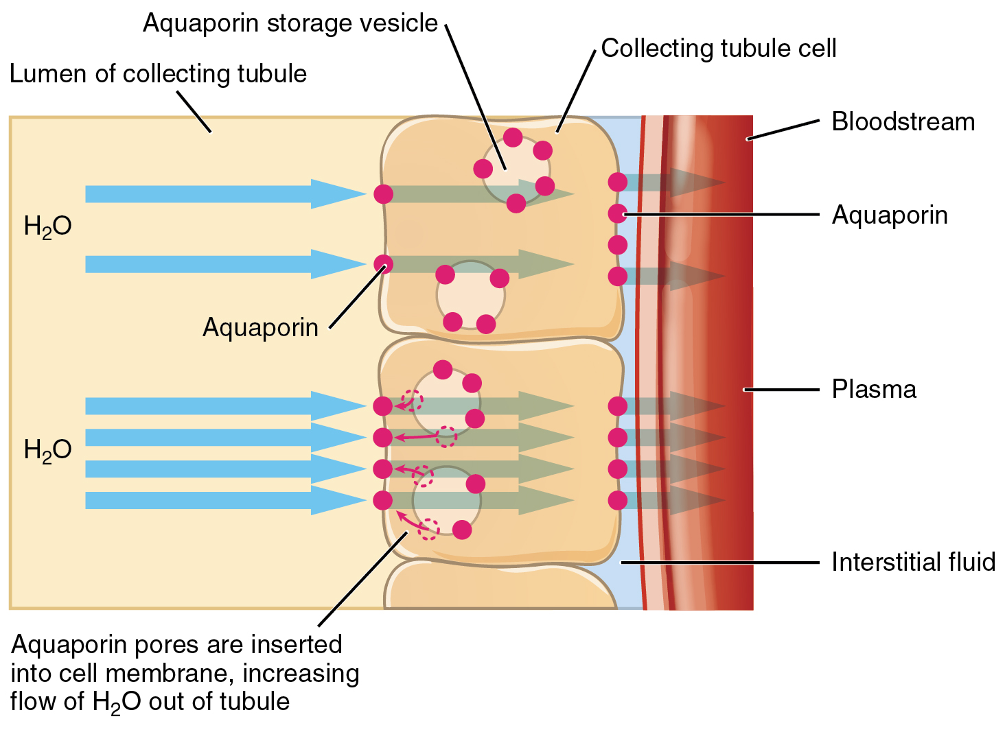
Diagram showing ADH-dependent insertion of aquaporin channels into collecting duct principal cells. Water moves osmotically from the tubular lumen, through aquaporins, to the interstitium and peritubular capillaries, concentrating the urine. This directly visualises the hormonal control step in osmoregulation. Source.
The duct walls are lined with principal cells, which respond to ADH by altering their permeability to water.
Mechanism of ADH Action
ADH binds to receptors on the cell surface membrane of the collecting duct epithelial cells.
This activates the enzyme adenylate cyclase, converting ATP to cyclic AMP (cAMP)—a secondary messenger within the cell.
cAMP triggers a cascade of events leading to the fusion of vesicles containing aquaporins with the cell membrane.
Aquaporins (water channel proteins) are inserted into the membrane, increasing its permeability to water.
Water moves out of the tubular fluid into the interstitial fluid and then into the capillaries surrounding the nephron by osmosis.
As a result, less water remains in the urine, producing small volumes of concentrated urine.
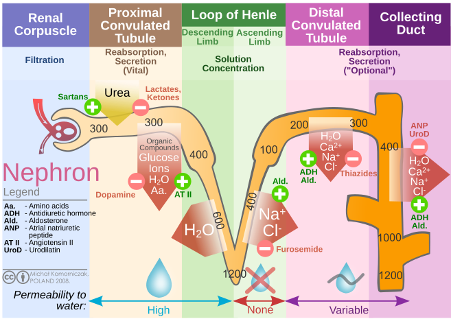
Overview of the nephron indicating urine osmolarity values along the tubule when ADH acts, culminating in a concentrated urine in the collecting duct. This contextualises the collecting-duct effect within the whole nephron. Extra detail included: segments of the nephron and transport processes not required by OCR may be shown. Source.
Aquaporins: Channel proteins embedded in cell membranes that facilitate the passive movement of water molecules along a water potential gradient.
When blood water potential rises again (due to rehydration), less ADH is secreted. The aquaporins are removed from the membrane by endocytosis, making the collecting duct walls less permeable to water, leading to dilute urine.
Summary of ADH Effects
Increased ADH: More aquaporins inserted, more water reabsorbed, concentrated urine produced.
Decreased ADH: Fewer aquaporins, less water reabsorbed, dilute urine produced.
This dynamic control allows the kidneys to precisely regulate body water content and maintain osmotic balance.
Integration with Other Homeostatic Mechanisms
The control of blood water potential operates as part of a negative feedback system within homeostasis.
Stimulus: Change in blood water potential (increase or decrease).
Receptor: Osmoreceptors in the hypothalamus detect the change.
Coordinator: Hypothalamic neurosecretory cells trigger or inhibit ADH release from the posterior pituitary.
Effector: Kidney collecting ducts alter water permeability.
Response: Water potential returns to normal, stabilising the internal environment.
Negative Feedback: A control mechanism where a change from the norm initiates responses that counteract the deviation, restoring equilibrium.
This feedback ensures stability: if water potential drops, ADH increases water reabsorption; if it rises, ADH secretion decreases. The system thus protects cells from osmotic stress.
Physiological Context and Significance
Maintaining correct blood water potential is essential for:
Cellular integrity: Preventing swelling or crenation of cells.
Blood pressure regulation: Influenced by plasma volume and osmotic pressure.
Efficient excretion: Concentration of nitrogenous waste without excessive water loss.
Thermoregulation support: Adequate hydration allows for effective sweating and evaporative cooling.
Disruption of ADH secretion or action leads to clinical conditions such as diabetes insipidus, characterised by excessive urination and dehydration due to failure in water reabsorption.
Through this tightly regulated hormonal mechanism involving osmoreceptors, the hypothalamus, the posterior pituitary, and ADH, the body maintains an optimal blood water potential, ensuring metabolic efficiency and homeostatic balance.
Practice Questions
Question 1 (2 marks)
Explain the role of osmoreceptors in the control of blood water potential.
Mark Scheme:
(1 mark) Osmoreceptors are specialised sensory neurones in the hypothalamus that detect changes in blood water potential.
(1 mark) When blood water potential decreases, osmoreceptors shrink, stimulating neurosecretory cells to release antidiuretic hormone (ADH) from the posterior pituitary.
Question 2 (5 marks)
Describe and explain how antidiuretic hormone (ADH) controls water reabsorption in the kidneys during dehydration.
Mark Scheme:
(1 mark) ADH is released from the posterior pituitary when osmoreceptors detect a decrease in blood water potential.
(1 mark) ADH binds to receptors on collecting duct cells in the nephron.
(1 mark) This activates a second messenger system (cAMP) inside the cell.
(1 mark) cAMP causes vesicles containing aquaporins to fuse with the cell surface membrane, inserting water channel proteins.
(1 mark) This increases the permeability of the collecting duct walls to water, allowing more water to be reabsorbed into the bloodstream, producing small volumes of concentrated urine.
FAQ
Osmoreceptors respond to changes in the osmotic pressure of the blood plasma surrounding them.
When blood water potential decreases (becomes more negative), water moves out of the osmoreceptor cells by osmosis, causing them to shrink. This physical change alters the membrane potential of the neurones, increasing their rate of nerve impulse firing.
Conversely, when blood water potential increases, water enters the osmoreceptors, making them swell and reducing nerve impulse frequency. This fine-tuned response allows the hypothalamus to adjust ADH release precisely.
ADH is a peptide hormone, meaning it is composed of short chains of amino acids rather than being lipid-based.
Because peptide hormones are hydrophilic, ADH cannot cross the phospholipid bilayer of target cell membranes. Instead, it binds to cell surface receptors on collecting duct cells.
This binding triggers an intracellular second messenger cascade involving cyclic AMP (cAMP), which ultimately leads to the insertion of aquaporins. This indirect mechanism ensures a rapid yet reversible response to water potential changes.
When ADH secretion decreases, the collecting duct cells reverse the process that inserted aquaporins.
The aquaporins are removed from the cell membrane by endocytosis, forming vesicles again within the cytoplasm.
The membrane then becomes less permeable to water, reducing water reabsorption.
As a result, urine becomes more dilute and is produced in greater volume.
This dynamic regulation ensures that the kidneys can adapt continuously to hydration status without permanent structural changes.
Alcohol inhibits the secretion of ADH from the posterior pituitary gland.
When ADH levels drop:
The collecting ducts become less permeable to water, so less water is reabsorbed.
This leads to the production of large volumes of dilute urine, causing dehydration
Dehydration in turn reduces blood water potential, often contributing to hangover symptoms such as headaches and fatigue.
This illustrates how external substances can disrupt homeostatic control mechanisms by interfering with hormonal signalling.
Although both hormones influence fluid balance, they act through different mechanisms and targets.
ADH controls water reabsorption by altering collecting duct permeability.
Aldosterone, a steroid hormone from the adrenal cortex, regulates sodium and potassium ion balance in the distal tubule and collecting duct.
Aldosterone promotes sodium reabsorption and potassium secretion, which indirectly increases water reabsorption by osmosis.
Thus, while ADH directly adjusts water permeability, aldosterone modifies solute movement that subsequently affects water balance.
