
OCR Specification focus:
‘Explain how regulated mitosis and programmed cell death shape body form, and how these processes respond to internal and external stimuli.’
Mitosis and apoptosis are vital cellular processes that ensure the correct growth, repair, and development of multicellular organisms. Controlled coordination between cell division and cell death shapes tissues, organs, and body structures throughout development.
The Role of Mitosis in Development
Mitosis is the process of nuclear division that produces two genetically identical daughter cells. It is central to growth, tissue repair, and the maintenance of genetically stable cells.
The Cell Cycle and Mitosis
The cell cycle consists of interphase (G₁, S, G₂) and mitosis (prophase, metaphase, anaphase, telophase), followed by cytokinesis. Regulation at checkpoints ensures accurate DNA replication and division.
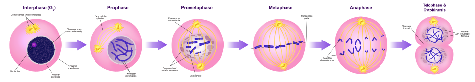
A labelled sequence of mitosis shows chromosome condensation, alignment at the metaphase plate, chromatid separation, and nuclear reformation, followed by cytokinesis. This visual supports how regulated mitosis increases cell number during development. Labels are limited to the stage names to maintain clarity. Source.
Mitosis: A type of cell division that results in two genetically identical daughter cells, each with the same number of chromosomes as the parent cell.
Key control points include:
G₁ checkpoint – ensures DNA is undamaged before replication.
G₂ checkpoint – checks successful DNA replication.
Spindle checkpoint (metaphase) – ensures chromosomes are correctly aligned before separation.
Cyclins and cyclin-dependent kinases (CDKs) regulate these transitions by activating or inhibiting key enzymes involved in DNA replication and mitosis.
Importance in Growth and Differentiation
During development, mitosis increases cell number, while cell differentiation leads to specialised tissues. Together, they establish the body’s form and function.
In the embryo, rapid mitotic divisions occur during early cleavage stages.
In later stages, controlled mitosis enables the growth of organs and tissues in response to genetic and environmental signals.
Stem cells divide by mitosis to provide progenitor cells that differentiate into specialised cell types.
Regulation of Mitosis
The precise regulation of mitosis prevents uncontrolled cell proliferation, which can lead to tumours. Internal control mechanisms include:
Proto-oncogenes, which promote mitosis when active.
Tumour suppressor genes, which inhibit cell division or trigger apoptosis when DNA damage occurs.
External signals, such as growth factors, nutrient availability, and hormonal cues, also modulate mitotic rates to align development with environmental conditions.
The Role of Apoptosis in Development
Apoptosis, or programmed cell death, is a genetically controlled process that removes unwanted or damaged cells. It is essential in shaping body structures and maintaining tissue health.
Apoptosis: A controlled sequence of events leading to the self-destruction and removal of a cell without causing inflammation or damage to neighbouring cells.
Mechanism of Apoptosis
Apoptosis involves a cascade of intracellular events, triggered by internal or external signals, leading to the activation of caspases—proteolytic enzymes that dismantle cellular components.
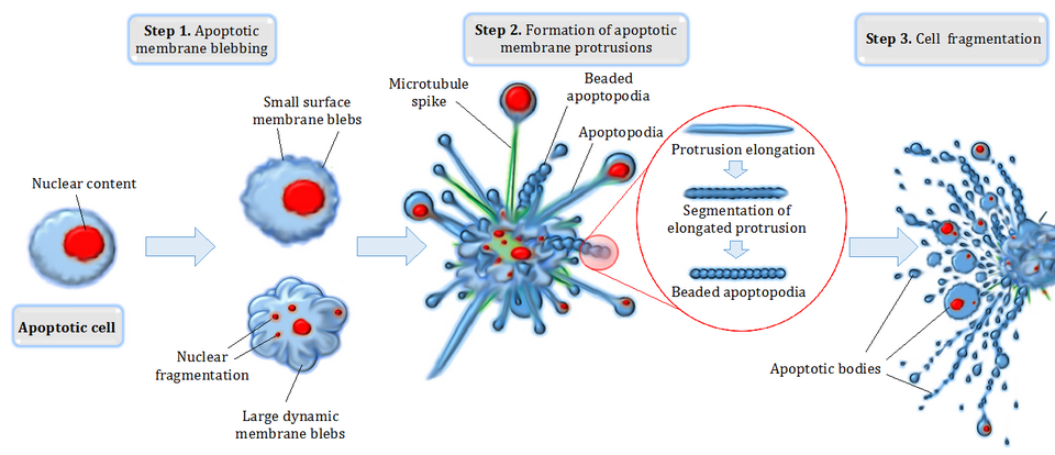
Diagram summarising apoptotic cell disassembly: shrinkage, blebbing, formation of apoptotic bodies, and their clearance. This underpins how apoptosis removes cells without inflammation, aiding tissue sculpting during development. The figure focuses on morphology; pathway proteins (e.g. Bcl-2 family) are not included, keeping it OCR-appropriate. Source.
Key stages include:
Cell shrinkage and condensation of chromatin.
Membrane blebbing, forming apoptotic bodies.
Phagocytosis of apoptotic bodies by macrophages or neighbouring cells.
This ensures that cell contents are safely recycled, maintaining tissue integrity.
Internal and External Triggers
Apoptosis may be initiated by:
Intrinsic signals: DNA damage, oxidative stress, or disruption of the mitochondrial membrane.
Extrinsic signals: Binding of death ligands to receptors such as Fas on the cell surface.
Developmental cues: Hormonal changes or growth factor withdrawal during specific stages of differentiation.
The Bcl-2 family of proteins regulates mitochondrial permeability, determining whether the apoptotic pathway proceeds. A balance between pro-apoptotic (e.g. Bax) and anti-apoptotic (e.g. Bcl-2) members ensures appropriate cell survival or death.
Coordinated Roles of Mitosis and Apoptosis
Development requires a precise equilibrium between cell proliferation and cell death. Too much mitosis without apoptosis results in overgrowth, while excessive apoptosis causes tissue loss.
Examples in Development
Embryonic limb formation: Cells between developing digits undergo apoptosis, creating distinct fingers and toes.
Nervous system development: Surplus neurons formed by mitosis are later removed by apoptosis, ensuring efficient synaptic connections.
Immune system maturation: Self-reactive lymphocytes are eliminated by apoptosis to prevent autoimmune disease.
Through such processes, organisms achieve accurate morphogenesis—the creation of body shape and organisation.
Molecular Control and Signalling Pathways
Cellular responses to developmental signals rely on signal transduction pathways that regulate gene expression and enzyme activity.
Intracellular Signalling
External stimuli such as hormones or growth factors activate receptor-mediated pathways involving second messengers like cyclic AMP (cAMP) and calcium ions (Ca²⁺).
These influence transcription factors that alter the expression of genes governing mitosis or apoptosis.
Checkpoint and Damage Responses
Cells with irreparable DNA damage activate the p53 protein, which halts the cell cycle or initiates apoptosis.
This prevents propagation of mutations and maintains genetic stability across generations of cells.
p53 Protein: A tumour suppressor that detects DNA damage and induces cell cycle arrest or apoptosis to prevent mutation accumulation.
Environmental and Hormonal Influences
External conditions can influence both mitotic and apoptotic activity.
Environmental Factors
Temperature and nutrient levels affect enzyme function and metabolic rates, altering mitotic activity.
Toxins or radiation can induce apoptosis through DNA damage pathways.
Hormonal Control
Hormones act as powerful regulators:
Thyroxine triggers apoptosis of tadpole tail cells during metamorphosis.
Oestrogen and growth hormone modulate mitotic activity during tissue development and repair.
These influences ensure developmental processes are responsive to both internal homeostatic cues and external environmental stimuli.
Importance in Health and Disease
Balanced mitosis and apoptosis are vital for normal development and lifelong maintenance.
Excessive mitosis can lead to cancer, where cells divide uncontrollably.
Insufficient mitosis results in growth disorders or delayed tissue repair.
Defective apoptosis contributes to tumour formation and autoimmune diseases, while excessive apoptosis can lead to neurodegenerative conditions.
Together, mitosis and apoptosis maintain cellular homeostasis, enabling organisms to grow, develop, and adapt safely to changing internal and external conditions.
Practice Questions
Question 1 (2 marks)
Describe one way in which apoptosis contributes to the normal development of an organism.
Mark scheme:
Award 1 mark for identifying a relevant developmental process (e.g. separation of digits in limbs, removal of surplus neurons).
Award 1 mark for explaining the effect of apoptosis on shaping body structure or function (e.g. removes cells between developing digits to form separate fingers and toes).
Question 2 (5 marks)
Explain how the regulation of mitosis and apoptosis ensures normal growth and development, and describe what might happen if this regulation fails.
Mark scheme:
1 mark: States that mitosis increases cell number for growth and repair.
1 mark: States that apoptosis removes damaged, aged or unnecessary cells to shape tissues.
1 mark: Explains that both processes are tightly regulated by genes and signalling pathways (e.g. proto-oncogenes, tumour suppressor genes, growth factors).
1 mark: Describes that a balance between mitosis and apoptosis maintains tissue size and structure (cellular homeostasis).
1 mark: Describes consequences of regulatory failure — e.g. excessive mitosis leading to tumours/cancer, or excessive apoptosis causing tissue loss or degenerative disease.
FAQ
Caspases are a family of protease enzymes that act in a cascade to dismantle the cell from within.
Initiator caspases are activated first, often by signals from mitochondria or cell-surface receptors.
These then activate executioner caspases, which cleave proteins in the cytoskeleton and nuclear envelope.
The result is cell shrinkage, DNA fragmentation, and membrane blebbing, forming apoptotic bodies that are removed by phagocytes.
This controlled breakdown ensures the cell’s contents do not leak, preventing inflammation.
Apoptosis is programmed cell death, whereas necrosis is accidental cell death due to injury or stress.
Apoptosis is regulated, energy-dependent, and involves orderly cell dismantling.
Necrosis results in cell swelling, rupture, and inflammation of surrounding tissue.
Apoptosis benefits the organism by removing specific cells, while necrosis is typically harmful and unplanned.
Growth factors are extracellular signalling molecules that stimulate mitosis and inhibit apoptosis in specific cells.
They bind to cell-surface receptors, triggering intracellular signalling cascades (often via kinase pathways).
These cascades lead to gene expression changes promoting cell-cycle progression and survival.
When growth factors are withdrawn, the lack of stimulation can activate pro-apoptotic pathways.
This balance allows tissues to grow when needed and limits cell numbers once development stabilises.
Mitochondria release key molecules that determine whether a cell will survive or undergo apoptosis.
When mitochondria are damaged or stressed:
They release cytochrome c into the cytoplasm.
Cytochrome c activates caspase-9, initiating the intrinsic apoptotic pathway.
Bcl-2 family proteins regulate this process by controlling mitochondrial membrane permeability.
Maintaining mitochondrial stability prevents unnecessary cell death and protects developing tissues.
Apoptosis maintains immune health by removing unneeded or harmful immune cells.
During immune development, self-reactive lymphocytes are eliminated to prevent autoimmune disease.
After an infection, activated immune cells die by apoptosis once their job is done, preventing excessive inflammation.
Phagocytic clearance of apoptotic cells also releases anti-inflammatory cytokines, maintaining immune balance.
This controlled turnover ensures immune precision without damaging healthy tissue.
