
OCR Specification focus:
‘Explain production of X-ray photons in the tube and factors affecting their energies.’
X-rays are produced when high-energy electrons interact with a metal target inside an X-ray tube, creating photons whose energies depend on accelerating voltage and target material.
Production of X-rays in the Tube
An X-ray tube is designed to convert the kinetic energy of fast-moving electrons into X-ray photons. This conversion occurs through interactions between the electrons and the atoms of a high-atomic-number target metal, typically tungsten, due to its high melting point and ability to withstand intense heating. Understanding these processes is essential for appreciating how the tube output is controlled and how different operating factors influence the resulting X-ray spectrum.
Electron Acceleration and Target Interaction
Electrons originate from a heated filament, where thermionic emission releases free electrons. These electrons are then accelerated across a large potential difference towards the anode. Their kinetic energy on reaching the target depends directly on this accelerating voltage.
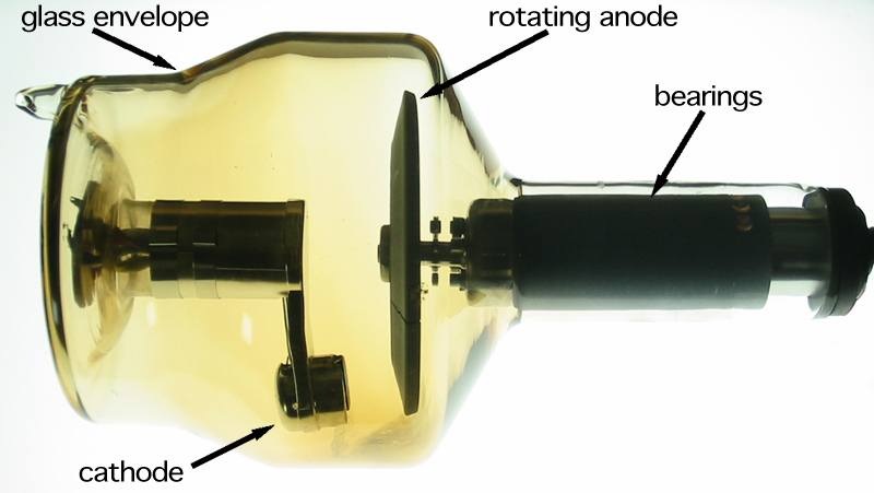
Diagram of a rotating-anode X-ray tube showing the heated cathode filament, focusing cup, evacuated glass envelope, tungsten anode, and the emerging X-ray photon beam. The image helps visualise how electrons are accelerated across the tube and focused onto the target to produce X-ray photons. Extra detail such as the rotor and stator for anode rotation is included but is not required by the OCR specification. Source.
Thermionic Emission: The release of electrons from a heated metal surface when thermal energy overcomes the binding forces holding electrons within the metal.
Once the electrons strike the metal target, they undergo rapid deceleration and numerous collisions. Only a small fraction of their kinetic energy is transformed into X-ray photons; the majority becomes thermal energy. The X-ray production process consists of two primary mechanisms: bremsstrahlung radiation and characteristic radiation. Each produces photons of different energies and contributes distinct features to the output spectrum.
A wide range of energies is possible, and their distribution depends on how the electron beam interacts with the target lattice. These interactions ensure that medical X-ray tubes emit a continuous spectrum with superimposed sharp peaks corresponding to characteristic emissions.
Bremsstrahlung (Braking Radiation)
Bremsstrahlung constitutes the dominant mechanism in most diagnostic tubes. As electrons decelerate in the electric field of target nuclei, they lose energy, which is emitted as X-ray photons. The amount of energy lost varies per event.
This means that bremsstrahlung photons form a continuous spectrum extending from very low photon energies up to a maximum value determined by the accelerating voltage.
Characteristic Radiation
Characteristic radiation arises from electron–atom interactions that eject inner-shell electrons from target atoms. When an electron from a higher energy level fills the vacancy, a photon with a discrete energy equal to the difference between the two levels is emitted. These sharp energy peaks appear at characteristic positions determined by the target material.
Characteristic Radiation: X-ray emission produced when an inner-shell electron is removed and an outer electron transitions to fill the vacancy, releasing a photon with a discrete energy.
These photons contribute to the line spectrum superimposed upon the bremsstrahlung background and ensure that changing the target material alters the energy positions of these peaks.
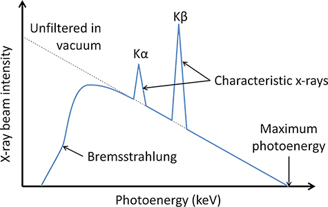
Graph of an X-ray energy spectrum showing a broad Bremsstrahlung distribution with superimposed sharp characteristic peaks at particular photon energies. The figure illustrates how most photons form a continuous range of energies while a smaller number occur at discrete energies determined by the target material. The graph includes extra detail on relative intensities beyond what is explicitly required in the OCR specification, but this supports deeper understanding. Source.
Factors Affecting X-ray Photons and Their Energies
Accelerating Voltage (Tube Potential)
The accelerating voltage is the most significant factor influencing X-ray photon energy. A higher voltage increases the kinetic energy of the electrons striking the target and therefore increases the maximum possible photon energy.
EQUATION
—-----------------------------------------------------------------
Maximum Photon Energy (E_max) = eV
E_max = Maximum energy of an emitted photon, measured in joules or electronvolts
e = Elementary charge, measured in coulombs
V = Accelerating potential difference, measured in volts
—-----------------------------------------------------------------
A higher voltage also increases the intensity of higher-energy photons within the bremsstrahlung distribution, shifting the spectrum towards shorter wavelengths.
Filament Current and Electron Number
The filament current controls the rate of thermionic emission and therefore the number of electrons in the beam. Increasing filament current raises the X-ray intensity but does not affect the maximum photon energy, as this depends solely on accelerating voltage.
Following changes in filament current, the tube output increases proportionally, meaning that more bremsstrahlung and characteristic photons are produced while maintaining the same energy distribution.
Target Material
Target material influences the energy of characteristic peaks and the efficiency of X-ray production. A higher atomic number increases the likelihood of bremsstrahlung interactions due to stronger nuclear electric fields. Tungsten is preferred for its durability and high atomic number, while molybdenum is sometimes used in specialised imaging such as mammography.
Tube Current and Exposure Duration
The tube current is the flow of electrons across the tube per second. Increasing tube current increases the total number of photons emitted, enhancing overall intensity without altering the energy of individual photons. Similarly, exposure duration affects the total photon count and is crucial for practical imaging control.
Control of X-ray Output
To achieve consistent and medically useful imaging, several parameters are carefully regulated:
Accelerating voltage (kV) — controls photon energy and penetration ability.
Tube current (mA) — sets the number of emitted photons.
Exposure time — determines total output.
Target material — influences efficiency and characteristic energies.
Tube filtration — removes low-energy photons that would increase patient dose without improving image quality.
Together, these factors govern the quality and utility of the X-ray beam, ensuring that diagnostic images are clear, controlled, and clinically effective.
Practice Questions
Question 1 (2 marks)
Electrons in an X-ray tube are accelerated towards a tungsten target by a high potential difference.
Explain why increasing the accelerating voltage increases the maximum energy of the X-ray photons produced.
Question 1 (2 marks)
Increasing the accelerating voltage increases the kinetic energy of the electrons striking the target. (1)
Higher-energy electrons can produce X-ray photons with greater maximum photon energy during deceleration. (1)
Question 2 (5 marks)
Describe how both bremsstrahlung radiation and characteristic radiation are produced in an X-ray tube.
Explain why bremsstrahlung produces a continuous spectrum while characteristic radiation produces discrete energy peaks.
Question 2 (5 marks)
Bremsstrahlung radiation: produced when electrons decelerate in the electric field of the target nuclei. (1)
Energy lost during deceleration is emitted as X-ray photons. (1)
Characteristic radiation: produced when an incoming electron ejects an inner-shell electron from a target atom. (1)
An outer electron transitions to fill the vacancy, emitting a photon with energy equal to the difference in energy levels. (1)
Bremsstrahlung produces a continuous spectrum because electrons lose variable amounts of energy during deceleration, while characteristic radiation has discrete energies determined by fixed atomic energy levels. (1)
FAQ
Tungsten has a very high atomic number (Z = 74), which increases the likelihood of bremsstrahlung interactions and makes X-ray production more efficient.
Its melting point is extremely high, allowing the target to withstand the intense heat generated when electrons decelerate.
Tungsten also has good thermal conductivity, helping to dissipate heat and reduce damage to the anode surface.
Filtration removes low-energy photons that contribute to patient dose without improving image quality.
• Aluminium filters preferentially absorb photons with energies below around 20–30 keV
• The resulting spectrum shifts towards higher average energies
• Beam becomes “harder”, meaning it has greater penetrating power
This modifies the usable output without altering the fundamental production mechanisms in the tube.
The focusing cup surrounds the filament and uses a negative potential to direct emitted electrons into a narrow beam.
This creates a small focal spot on the anode, ensuring sharper image resolution.
A well-focused beam also increases the intensity of X-ray production at the target by preventing electrons from spreading out and striking a wider area.
A rotating anode spreads heat over a larger surface area, reducing the risk of target damage.
• Allows much higher tube currents than stationary anodes
• Enables longer exposure times
• Reduces pitting and wear on the target surface
• Improves tube lifespan and consistency of X-ray output
This is essential for producing high-quality diagnostic images at clinically useful intensities.
Conversion efficiency is low—usually less than 1%—but it varies with several parameters.
• Higher accelerating voltages increase efficiency because electrons interact more strongly with target nuclei
• Higher atomic number targets generate more bremsstrahlung events
• Smaller focal spots concentrate energy, increasing local photon production
Despite these factors, most energy is always converted into heat, requiring robust cooling and anode design.

.jpg)