
OCR Specification focus:
‘Describe CAT scanner components and explain advantages over single X-ray images.’
Computed Axial Tomography (CAT scanning) uses rotating X-ray sources and sophisticated computer reconstruction to create detailed cross-sectional images, revealing internal structures with clarity far beyond a single X-ray image. This technique significantly improves diagnostic accuracy and enables clinicians to distinguish tissues that would otherwise overlap in conventional radiography.
CAT Scanner Components
The operation of a CAT scanner relies on coordinated mechanical, electronic, and computational systems arranged to gather multiple X-ray projections around the patient.
Gantry and Rotating X-ray Assembly
The gantry is the circular frame through which the patient passes. It houses the rotating X-ray tube, which emits a narrow, highly collimated X-ray beam.
The tube rotates rapidly (typically several revolutions per second).
X-ray photons pass through the patient from many angles.
Rotation reduces motion blur and increases sampling density.
As the X-ray tube rotates, the beam continuously irradiates a thin slice of the patient’s body, enabling the scanner to accumulate projection data needed for image reconstruction.

A schematic illustration showing an X-ray tube and detector array rotating around the patient to acquire many projections for CT reconstruction. Source.
Detector Array
Opposite the X-ray tube lies a curved or ring-shaped detector array, consisting of hundreds to thousands of sensitive detectors.
Detectors measure intensity reductions caused by attenuation through tissues.
Modern scanners often use scintillation detectors coupled to photodiodes for fast, accurate photon detection.
The large number of detectors ensures comprehensive angular coverage.
The system records how much of the X-ray beam is absorbed along each path, producing a set of attenuation profiles.
Patient Table and Positioning System
The patient rests on a motorised table that moves smoothly through the gantry.
The movement is precise to ensure that slices correspond accurately to anatomical planes.
In helical (spiral) scanning, the table moves continuously as the tube rotates, generating a helical dataset.
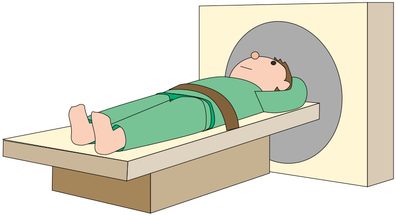
An illustration of a patient positioned on a motorised table moving into the circular gantry that houses the rotating X-ray tube and detectors. Source.
Computer System
A powerful computer controls the tube rotation, detector acquisition, and preprocessing of raw data.
The computer applies mathematical reconstruction algorithms, typically based on filtered back-projection or iterative reconstruction.
It corrects for noise, beam hardening, and detector inconsistencies.
Final images are displayed as greyscale slices representing different attenuation values.
Image Formation in CAT Scanning
The core idea is that each detector reading represents the cumulative attenuation along a single path. By collecting many such readings from multiple angles, the scanner can reconstruct the internal density distribution slice by slice.
Pixel Values and Tissue Differentiation
Each pixel in a reconstructed slice corresponds to a tiny volume called a voxel. The voxel is assigned a numerical value representing linear attenuation coefficient for that region.
Linear attenuation coefficient: A measure of how strongly a material absorbs or scatters X-rays per unit distance.
These values are typically converted into Hounsfield units, which standardise tissue contrast and allow clinicians to distinguish between air, fat, soft tissue, bone, and contrast-enhanced regions.
Between definition blocks, it is helpful to understand that tissue-specific attenuation values allow CAT scanning to surpass conventional radiography, where overlapping structures obscure detail.
Advantages of CAT Scanning Over Single X-ray Images
CAT scanning provides significant improvements in clarity, diagnostic detail, and clinical utility.
Elimination of Superposition
Conventional radiographs compress three-dimensional anatomy into a two-dimensional projection, producing overlapping shadows. CAT scanning removes this limitation.
Each slice isolates a thin section of tissue.
Structures are no longer hidden behind others.
Pathologies such as tumours or bleeding become easier to detect.
High Contrast Resolution
CAT scanners differentiate tissues with small differences in attenuation.
Tumours, cysts, or soft-tissue irregularities may be invisible in a standard X-ray but clearly visible in CAT images.
Computer reconstruction enhances subtle contrast variations.
Three-Dimensional Reconstruction
Sequential slices can be combined into full 3D models.
Useful for surgical planning and navigation.
Enables volumetric measurements of organs or lesions.
Improves visualisation of complex anatomical regions, especially the head, chest, and abdomen.
Rapid and Non-invasive
Modern CAT scanners acquire whole-body images in seconds.
Particularly valuable in trauma assessment.
Fast scanning reduces movement artefacts and increases diagnostic accuracy.
Non-invasive nature makes it suitable for repeated monitoring.
A significant practical advantage is that CAT scanning often requires lower operator interpretation skill for complex regions because reconstructed slices present information more intuitively than overlapping radiographs.
Use of Contrast Agents
Although not mandatory, CAT scans often employ iodine-based contrast media to distinguish between blood vessels, soft tissues, and pathological developments.
Enhances imaging of vascular structures.
Highlights perfusion abnormalities.
Improves tumour visibility.
Normal sentences between structured blocks maintain readability, and it is worth noting that contrast-enhancement complements the intrinsic resolution of the CAT scan, further improving diagnostic value.
Quantitative Data for Diagnosis
CAT scanning allows measurement of attenuation values and tissue densities.
Enables tracking of disease progression.
Provides objective data for medical decision-making.
Supports detailed analysis of bone, lung tissue, and blood flow patterns.
Summary of Key Advantages
CAT scanning offers a combination of structural clarity, high resolution, rapid acquisition, and computational precision unavailable with single X-ray images.
Layered, slice-based imaging without overlapping anatomy
Exceptional contrast resolution
3D reconstruction capabilities
Quick imaging suitable for emergencies
Improved diagnostic accuracy through quantifiable image data
Practice Questions
(1–3 marks)
State two main components found inside the gantry of a CAT scanner and briefly describe their roles in image formation.
Question 1 (1–3 marks)
Award 1 mark for each correct component (max 2 marks), and 1 mark for an appropriate description of role (max 1 mark).
Possible correct points:
X-ray tube – emits a narrow beam of X-rays that rotates around the patient.
Detector array – detects transmitted X-rays after they pass through the patient.
Collimator – shapes the X-ray beam (accept if mentioned correctly).
Gantry – houses the rotating assembly (only credit if described inside the gantry).
Descriptions that earn the third mark (any one of the following):
X-ray tube rotates to obtain many projections from different angles.
Detectors measure attenuated intensities to generate projection data.
Collimator ensures a thin, well-defined beam for slice imaging.
(4–6 marks)
Explain how a CAT scanner produces cross-sectional images of the body and why these images provide greater diagnostic detail than a single X-ray image. Your answer should refer to attenuation measurements, reconstruction, and superposition.
Question 2 (4–6 marks)
Award marks for the following valid physics points, up to 6 total:
Rotation of X-ray tube and detectors around the patient to collect multiple projections. (1 mark)
Measurement of attenuation of the X-ray beam by detectors for many different paths. (1 mark)
Computer reconstruction (e.g., filtered back-projection or similar wording) to form a cross-sectional slice. (1 mark)
Slices represent attenuation coefficients assigned to pixels/voxels. (1 mark)
Elimination of superposition compared with a single X-ray image. (1 mark)
Greater diagnostic detail/contrast resolution because tissues with small attenuation differences can be distinguished. (1 mark)
FAQ
In helical scanning, the patient table moves continuously through the gantry while the X-ray tube rotates, forming a spiral path of data around the body.
This allows faster image acquisition, reduces breathing artefacts, and enables high-quality 3D reconstructions because no gaps appear between slices.
Traditional step-and-shoot scanning pauses table movement for each slice, which increases scan time and is more prone to misalignment between slices.
High rotation speeds minimise motion blur caused by breathing, heartbeat, or patient movement.
They also increase the number of projection angles collected per unit time, improving spatial resolution and reducing noise in the reconstructed slices.
Modern scanners can complete a full rotation in under half a second, enabling near real-time imaging in emergency settings.
Resolution is limited by detector size, beam collimation, X-ray focal spot size, and patient movement.
CAT scanning relies on measuring attenuation along many paths, and reconstruction algorithms average data across voxels. This inherently limits fine detail.
Additionally, higher resolution requires higher radiation dose, so safety limits prevent pushing resolution indefinitely.
Iterative methods repeatedly refine an estimated image by comparing predicted projections with actual measured data.
They reduce noise, suppress artefacts, allow sharper edges, and can achieve good image quality at lower radiation doses.
Filtered back-projection is faster but more prone to streak artefacts, especially when attenuation differences are large or projections are limited.
Brightness corresponds to the tissue’s attenuation coefficient, which depends on density and atomic number.
For example:
Bone appears bright because it strongly attenuates X-rays.
Fat appears darker because it attenuates weakly.
Soft tissues fall in between.
This natural contrast enables identification of many structures, even before contrast enhancement is used.
