
OCR Specification focus:
‘Explain use of contrast agents such as barium and iodine for X-ray imaging.’
Contrast media enhance differences in X-ray absorption between soft tissues, allowing clearer visualisation of internal structures that would otherwise produce minimal contrast in radiographic images.
Contrast Media in X-ray Imaging
Contrast media are specialised substances introduced into the body to improve the visibility of soft tissues during X-ray procedures. Since many biological tissues have similar attenuation coefficients—meaning they absorb X-rays to similar degrees—the resulting images may lack distinguishable boundaries or sufficient diagnostic detail. Contrast agents overcome this by introducing materials with significantly different X-ray absorption characteristics, enabling clinicians to obtain clearer and more informative radiographs.
Why Soft Tissues Require Contrast Enhancement
Soft tissues such as blood vessels, intestines, or the renal system exhibit relatively low X-ray attenuation because they are composed of low-atomic-number elements. In a standard radiograph, these tissues appear with little distinction. To address this limitation, contrast agents containing high-atomic-number elements are used, as their atoms possess a large number of electrons capable of interacting strongly with incoming X-ray photons.
Interaction Mechanism of Contrast Agents with X-rays
The diagnostic effectiveness of contrast media arises from the photoelectric effect, a dominant attenuation mechanism when X-ray photon energies are comparable to the binding energies of inner-shell electrons in heavy atoms. High-Z elements such as iodine (Z = 53) and barium (Z = 56) have suitable electron energies to encourage this interaction.
The photoelectric effect probability scales approximately with Z3Z^3Z3, meaning even small increases in atomic number lead to dramatic increases in attenuation.
When a contrast medium fills or coats a structure, it causes that region to absorb many more X-rays than surrounding tissues.
On the resulting X-ray image, high-attenuation areas appear lighter or more opaque, clearly outlining the target anatomical feature.
This attenuation difference provides clinicians with the information needed to identify abnormalities, blockages, or structural irregularities.
Types of Contrast Media
Two major contrast media used in X-ray imaging are barium sulphate and iodine-based compounds, each suited to different diagnostic tasks.
Barium Sulphate
Barium sulphate is a radiopaque compound used primarily for imaging the gastrointestinal (GI) tract.
Radiopaque: A material that strongly absorbs X-rays and therefore appears light on a radiograph.
Barium sulphate is administered orally or rectally, depending on the region of interest. Its insolubility makes it safe for use within the digestive system because it is not absorbed into the bloodstream. After coating the internal lining of the oesophagus, stomach, or intestines, it creates a clear outline of these structures on an X-ray image.
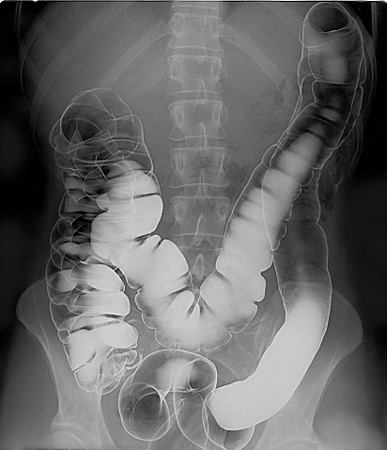
Double-contrast barium enema radiograph of the large intestine. The bright white regions show where barium sulphate has coated the intestinal lining, producing a sharply defined outline. Some labelled anatomical detail exceeds the syllabus requirement but remains helpful visual context. Source.
Iodine-Based Contrast Agents
Iodine compounds are typically injected into the bloodstream because they are water-soluble and can circulate through vascular structures.
Vascular system: The network of blood vessels through which blood circulates in the body.
These agents allow radiographers to visualise arteries, veins, and organs that take up iodine. Iodine-based media are commonly used in procedures such as angiography, intravenous urography, and CT scans requiring enhanced soft-tissue contrast.
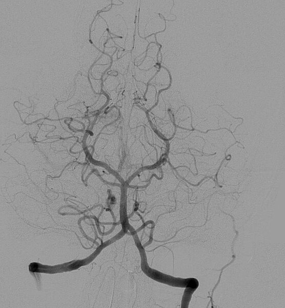
Cerebral angiogram showing arteries filled with an iodine-based contrast agent. The contrast makes the vascular tree stand out clearly against surrounding tissues. Fine vessel branching is visible, slightly beyond syllabus depth but valuable for understanding contrast enhancement. Source.
Normal circulation distributes the iodine throughout the body, but imaging is timed so that X-rays are taken while the medium is concentrated in the structure of interest. This timing makes dynamic processes—such as blood flow or renal filtration—visible on radiographs.
How Contrast Media Are Delivered
Contrast agents can be administered in several ways depending on the anatomical region:
Oral administration: Barium meals or barium swallow tests for oesophagus and stomach imaging.
Rectal administration: Barium enemas for lower-GI investigations.
Intravenous injection: Iodine-based media for vascular imaging, kidney studies, or CT contrast-enhanced scans.
Intra-arterial injection: Used in angiography to highlight specific arteries.
These delivery routes ensure the contrast medium reaches and outlines the correct structure while minimising systemic risk.
Diagnostic Uses of Contrast Media
Contrast media enable a range of medical investigations across multiple systems:
Gastrointestinal Tract
A barium swallow test can identify conditions such as tumours, ulcers, narrowing of passages, or motility disorders. Because the barium outlines the lumen, radiographers can easily observe any irregularities.
Blood Vessels
Iodine-based contrast makes arteries and veins visible, allowing detection of blockages, aneurysms, or malformations. Its rapid clearance from the bloodstream also enables sequential imaging of dynamic processes.
Kidneys and Urinary System
Intravenous urography uses iodine contrast to visualise renal filtration and urinary tract structures, providing insight into obstructions or functional abnormalities.
Soft Tissues with Poor Natural Contrast
Organs such as the liver or spleen may also be imaged with iodinated contrast to reveal lesions or tumours that would otherwise be difficult to detect.
Safety Considerations
Although contrast agents are generally safe, clinicians must assess potential risks:
Patients may experience mild side effects such as warmth or nausea following iodine injection.
Allergic-type reactions to iodine-based media are possible and must be monitored.
Barium sulphate is unsuitable when perforation of the GI tract is suspected; iodine-based alternatives are used instead.
Understanding these considerations ensures the safe and effective use of contrast media in X-ray imaging.
Practice Questions
Question 1 (2 marks)
Explain why barium sulphate is used as a contrast medium for imaging the gastrointestinal tract in X-ray procedures.
Question 1 (2 marks)
Barium sulphate strongly absorbs X-rays / has a high attenuation coefficient / is radiopaque. (1 mark)
It coats the lining of the gastrointestinal tract, allowing clear outlines to be seen on X-ray images. (1 mark)
Question 2 (5 marks)
A patient is injected with an iodine-based contrast agent before undergoing an angiography scan.
(a) Describe how the iodine-based contrast medium enhances the visibility of blood vessels in the resulting X-ray images.
(b) Explain why iodine is suitable for this role, referring to both its atomic properties and how X-rays interact with matter.
(c) State one possible safety consideration when using iodine-based contrast agents.
Question 2 (5 marks)
(a)
Iodine increases X-ray absorption in the blood vessels, making them appear more clearly / more opaque on the X-ray image. (1 mark)
This produces contrast between the vessels and surrounding soft tissues. (1 mark)
(b)
Iodine has a high atomic number, meaning many electrons for interaction with X-ray photons. (1 mark)
Increased likelihood of photoelectric effect / strong attenuation at diagnostic X-ray energies. (1 mark)
(c)
Possible allergic reaction or side effects such as warmth or nausea. (1 mark)
FAQ
High-atomic-number elements have tightly bound inner-shell electrons, which increases the probability of photoelectric absorption at diagnostic X-ray energies. This effect causes a significant rise in attenuation compared to surrounding soft tissues, creating clear image contrast.
Their strong interaction with X-rays also allows a lower quantity of contrast agent to produce a noticeable difference in opacity, making them both effective and efficient for clinical use.
Pure barium ions are toxic because they dissolve in bodily fluids, but barium sulphate is highly insoluble. This means it does not dissociate into harmful ions.
Because it remains chemically inert as it travels through the gastrointestinal tract, it passes through the body unchanged and is not absorbed into the bloodstream.
Visibility depends on how quickly the agent mixes with blood, its rate of uptake by organs, and renal clearance.
Key factors include:
Solubility and viscosity of the specific iodine formulation
The patient’s cardiac output, which affects circulation speed
Kidney function, which determines how fast the agent is filtered and excreted
Combined contrast enhances structural detail by simultaneously highlighting and surrounding an anatomical region with contrasting densities.
For example:
Barium sulphate may coat the inner surface of the intestines (positive contrast).
Air may be introduced to fill the lumen (negative contrast).
This double-contrast technique produces a sharper view of the mucosal lining and helps identify small abnormalities.
Iodine agents are water-soluble, allowing them to be rapidly distributed and eliminated through the kidneys.
Their formulations are designed to minimise viscosity and osmolarity so they move efficiently through blood vessels and reduce patient discomfort.
Although repeat imaging is common, clinicians still monitor cumulative exposure to ensure kidney function remains stable.
