
OCR Specification focus:
‘Describe PET: positron–electron annihilation and image formation from coincident gamma photons.’
Positron Emission Tomography (PET) provides functional medical images by detecting gamma photons produced when emitted positrons annihilate with electrons, enabling clinicians to study metabolic activity in tissues.
PET Scanner Physics
Radioactive decay and positron emission
PET imaging relies on radionuclides that decay through β⁺ emission, producing positrons with small kinetic energies suitable for medical imaging.
Positron: The antiparticle of the electron, identical in mass but carrying a positive charge.
Once emitted from the decaying nucleus, the positron travels a short distance through tissue. This distance, called the positron range, depends on the isotope’s energy and affects image resolution. Understanding this behaviour helps explain why PET images reveal function rather than just structure. After slowing down through interactions with surrounding electrons, the positron eventually encounters an electron.
Positron–electron annihilation
When a positron collides with an electron, they undergo annihilation, a process where their mass is converted entirely into energy. Two gamma photons are produced, each with an energy of 511 keV, and they travel in almost exactly opposite directions (approximately 180° apart). This predictable geometry is essential for reconstructing accurate PET images.
Annihilation radiation: A pair of gamma photons produced when a positron and electron mutually destroy each other, each carrying 511 keV of energy.
Between releasing the annihilation radiation and their detection by the PET scanner, the photons may undergo interactions such as Compton scattering. These interactions can degrade image quality, so scanners use systems of detectors and reconstruction algorithms to identify genuine photon pairs.
Coincidence detection
A defining feature of PET imaging is coincidence detection, a method used to ensure that only photon pairs originating from the same annihilation event are registered.
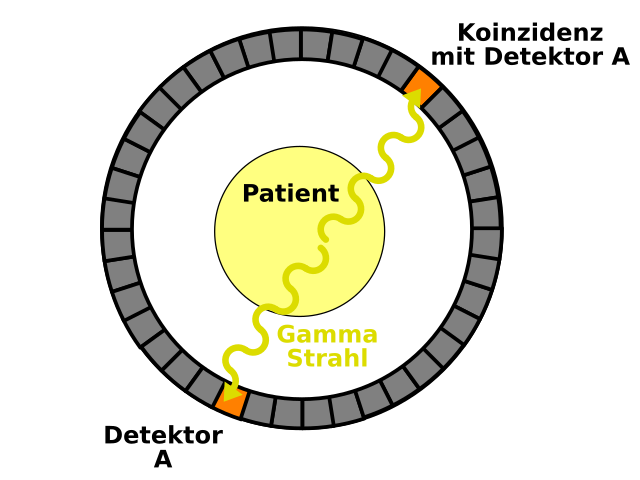
This diagram shows the principle of coincidence detection in PET: two gamma photons from a single annihilation event travel in opposite directions and are recorded simultaneously by two detectors. The line joining these detectors represents the line of response used in reconstructing the tracer distribution. Some labels are in German and include extra geometric detail beyond the OCR syllabus, but they do not affect the core physical idea. Source.
Each detector ring contains many individual detectors made from materials with high gamma absorption efficiency.
When two opposing detectors record gamma photons within a very short time window (typically a few nanoseconds), this simultaneous detection is termed a coincidence event.
The line joining the two detectors is defined as the line of response (LOR).
Repeated detection of many LORs originating from the distribution of annihilation events allows reconstruction of a 3D image.
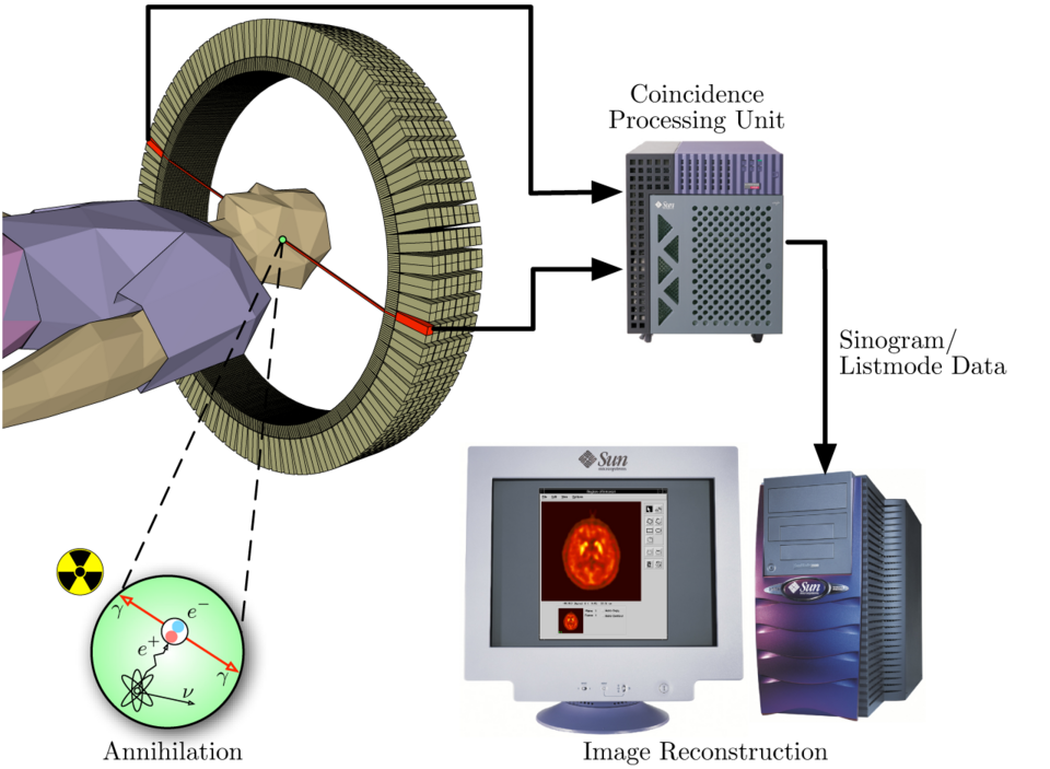
This schematic illustrates how a PET scanner detects pairs of gamma photons from annihilation events, forms lines of response, and reconstructs an image on a computer. It highlights the detector ring, coincidence processing unit, and image reconstruction stage. The specific scanner hardware and computer model shown are illustrative and not required by the OCR syllabus. Source.
This method avoids the need for physical collimators and increases efficiency compared with systems such as gamma cameras.
Components of a PET Scanner
Detector rings and scintillation crystals
PET scanners are built from circular or cylindrical rings of detectors containing scintillation crystals such as lutetium oxyorthosilicate (LSO) or bismuth germanate (BGO). These materials efficiently absorb high-energy gamma photons and emit light pulses in response.
Scintillation: The emission of light from a material when it absorbs ionising radiation.
The crystals are coupled to photodetectors, traditionally photomultiplier tubes (PMTs) but increasingly silicon photomultipliers (SiPMs), which convert the light into electrical signals. These signals provide precise timing information, allowing the system to determine when pairs of photons arrive almost simultaneously. Between this detection and the final display, several layers of processing refine the image and reduce noise.
Time-of-flight PET
Modern PET scanners often incorporate time-of-flight (TOF) information, which measures the tiny difference in arrival time between the two photons. This allows more accurate localisation of the annihilation event along the LOR.
TOF reduces background noise.
It improves spatial resolution.
It enhances image contrast, particularly useful for imaging larger patients.
Although TOF does not change the fundamental physical processes in PET, it greatly improves the final quality of diagnostic images.
PET Image Formation
Data acquisition and reconstruction
PET images are formed by combining thousands of coincidence events.
Each coincidence defines an LOR.
Many LORs crossing at the site of tracer concentration indicate high activity there.
Reconstruction algorithms such as filtered back projection and iterative reconstruction convert this raw data into a detailed 3D image.
Following reconstruction, the scanner’s computer assigns colour scales or intensity values to represent areas of higher or lower tracer concentration, allowing clinicians to interpret metabolic or functional activity clearly.
Functional imaging and tracer distribution
Unlike structural imaging (e.g., X-rays or CAT scans), PET visualises biochemical processes. PET radiotracers—commonly those linked to glucose analogues—accumulate in tissues in proportion to their metabolic rates. Regions with higher uptake generate more annihilation events and therefore produce more coincident gamma detections along intersecting LORs.
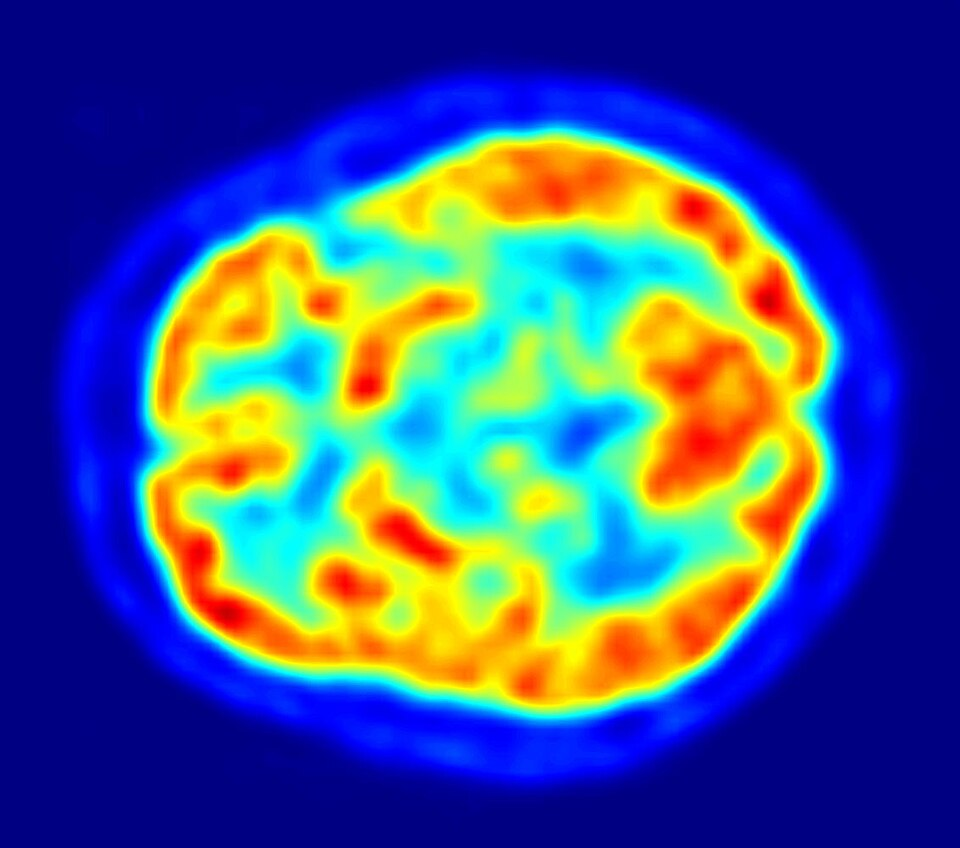
This PET image shows a horizontal slice through the human brain, with red and yellow regions indicating high uptake of the radiotracer and blue areas indicating low uptake. It reinforces that PET images display metabolic activity rather than anatomy. The source page references the tracer ¹⁸F-FDG and specific scanner details, which are additional contextual information not required by the OCR syllabus. Source.
This distribution enables PET to detect abnormalities in physiology, such as altered metabolism in tumours, even before structural changes appear. Thus, PET offers clinicians a powerful tool for early detection and precise mapping of disease.
This combination of positron emission, annihilation physics, and coincidence detection is central to understanding PET scanner operation and the formation of diagnostic images.
Practice Questions
Question 1 (2 marks)
A positron emitted inside the body travels a short distance before annihilating with an electron.
(a) State what is produced in this annihilation event.
(b) Explain why these products travel in nearly opposite directions.
Question 1
(a)
• Two gamma photons are produced. (1)
(b)
• Momentum is conserved in the annihilation event. (1)
• The electron and positron have negligible momentum at the instant of annihilation, so the photons travel in nearly opposite directions. (1)
(Max 1 mark for part (b))
Question 2 (5 marks)
Describe how a PET scanner forms an image from the gamma photons produced in positron–electron annihilation. In your answer, include:
• what happens at the site of annihilation
• how the photons are detected
• what is meant by coincidence detection
• how the final image is produced.
Question 2
Award marks for the following points, up to a maximum of 5:
• Positron from the tracer meets an electron and they annihilate. (1)
• Two gamma photons of energy 511 keV are emitted. (1)
• Photons travel in almost exactly opposite directions. (1)
• Detectors arranged in a ring detect the photons. (1)
• Coincidence detection: only pairs of photons arriving simultaneously at opposite detectors are counted. (1)
• Each coincidence defines a line of response along which the annihilation occurred. (1)
• A computer uses many such lines to reconstruct a 3D image showing tracer concentration. (1)
FAQ
The resolution partly depends on the positron range, which varies with the maximum positron energy of the isotope. Higher-energy positrons travel farther before annihilation, introducing greater uncertainty in where the event occurred.
Other factors include:
• the statistical rate of decay producing usable events
• how well the detectors measure timing information
• reconstruction algorithms that compensate for high-energy isotope behaviour
These materials have high atomic numbers, giving them strong gamma-photon stopping power essential for detecting 511 keV photons efficiently.
They also produce fast, intense light pulses, which:
• improve timing resolution for coincidence detection
• reduce signal overlap at high count rates
• enable time-of-flight (TOF) PET performance
TOF narrows the possible location of annihilation along each line of response by using the small difference in photon arrival times. This reduces the amount of “background” volume contributing to the reconstruction.
As a result:
• fewer incorrect locations are included in the image
• iterative algorithms converge more quickly
• contrast improves in regions with low tracer uptake
Some photons undergo Compton scattering in the body before reaching a detector. These scattered photons arrive from the wrong direction and lower the accuracy of coincidence events.
Scatter correction algorithms:
• estimate the fraction of scattered photons
• subtract or reweight affected data
• preserve accurate tracer distribution, especially in soft tissues
Random coincidences occur when two unrelated photons happen to hit opposite detectors within the time window.
PET scanners reduce this by:
• narrowing the coincidence timing window
• using high-speed electronics to reject mismatched signals
• implementing software corrections that estimate random rates from single-photon detections
This improves image fidelity by keeping only genuine annihilation events.
