
OCR Specification focus:
‘Describe tracers technetium-99m and fluorine-18 and their medical applications.’
Medical tracers enable clinicians to visualise physiological processes inside the body by detecting emitted radiation from suitably chosen radioactive isotopes. They play a crucial role in diagnostics.
The Purpose of Medical Tracers
Medical tracers are radioactive substances introduced into the body to track the movement, uptake, or concentration of materials in specific tissues. Their diagnostic value arises from the radiation they emit, which external detectors can measure to construct functional images. These images reveal abnormalities in organ function, tissue metabolism, and blood flow in ways structural imaging alone cannot achieve. Because tracers are used within living systems, careful choice of isotope is essential to ensure patient safety, minimise radiation dose, and achieve high-quality diagnostic information.
Key Properties of an Effective Medical Tracer
For a radioactive isotope to be suitable as a medical tracer, it must meet stringent physical and biological criteria. The two isotopes on the OCR specification — technetium-99m (Tc-99m) and fluorine-18 (F-18) — exemplify these requirements. Typical properties include:
Emission of gamma radiation (or annihilation photons) that can escape the body and be detected externally.
A short half-life to minimise patient dose while remaining long enough for imaging.
Chemical compatibility allowing the isotope to be incorporated into biologically relevant compounds.
Minimal biological toxicity.
These considerations guide why Tc-99m is dominant in nuclear medicine and why F-18 is central to PET imaging.
Technetium-99m as a Tracer
Technetium-99m is the most widely used tracer in gamma-camera and SPECT imaging due to its highly favourable nuclear properties and chemical versatility. The “m” denotes its metastable state, meaning it exists for a short time before decaying and emitting detectable gamma photons.
Metastable state: A temporarily excited nuclear state that persists long enough to be detected before decaying to a lower energy state.
Tc-99m decays by isomeric transition, emitting a gamma photon with energy well-suited for external detection. This allows imaging systems to form clear, high-contrast images with limited attenuation. Its short half-life (approximately six hours) ensures that residual radiation is low after the procedure while still providing time for clinical workflows.
Why Technetium-99m Is So Widely Used
Tc-99m can form compounds with a wide range of ligands, allowing it to target specific tissues. Key advantages include:
Low radiation dose to the patient due to short half-life.
No beta emission, making it safer for surrounding tissues.
High gamma photon yield improving detection efficiency.
Flexible chemistry, enabling the creation of many radiopharmaceuticals.
Applications of Technetium-99m in Medicine
Tc-99m is incorporated into pharmaceuticals tailored to different diagnostic tasks. Common medical applications include:
Bone scans — Tc-99m-labelled phosphates bind to bone tissue, highlighting fractures, tumours, and metabolic abnormalities.
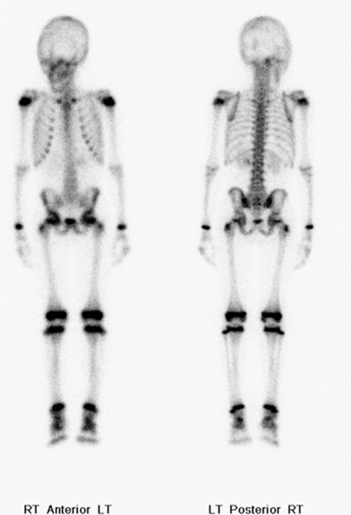
Whole-body bone scintigraphy using technetium-99m MDP shows expected tracer uptake throughout the skeleton and physiological excretion pathways. This illustrates how Tc-99m localises in bone tissue to reveal fractures, tumours, and metabolic abnormalities. The anatomical distribution shown goes beyond OCR requirements but reinforces clinical context. Source.
Kidney imaging — tracers such as Tc-99m-MAG3 assess kidney perfusion and filtration.
Cardiac imaging — Tc-99m-sestamibi is used to evaluate myocardial perfusion and detect ischaemic regions.
Thyroid imaging — Tc-99m-pertechnetate is taken up by the thyroid gland to detect nodules and assess function.
These varied applications arise from the isotope’s adaptable chemistry, which allows precise targeting of organs or biological pathways.
Fluorine-18 as a Tracer
Fluorine-18 is a positron-emitting isotope used primarily in positron emission tomography (PET). Its decay mechanism directly supports high-resolution, functional imaging through detection of coincident gamma photons.
Positron emission: A nuclear decay process in which a proton converts into a neutron and releases a positron, the antimatter counterpart of the electron.
After being emitted, the positron travels a short distance before annihilating with an electron. This produces two gamma photons travelling in opposite directions, which PET detectors record to reconstruct detailed images of tracer distribution. The half-life of F-18 (approximately 110 minutes) is short yet long enough to allow synthesis, transport, and imaging.
The Role of F-18 in PET Imaging
F-18 is typically incorporated into biologically active molecules. The most important example is fluorodeoxyglucose (FDG), a glucose analogue that accumulates in tissues with high metabolic activity. PET imaging with F-18-FDG reveals functional details inaccessible to purely structural imaging techniques.
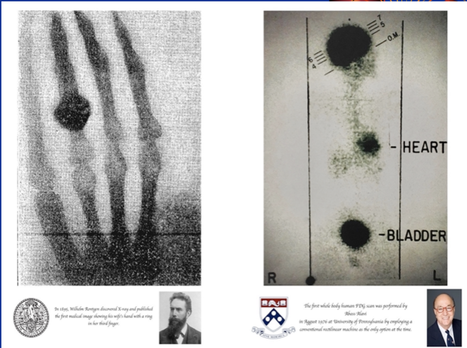
This figure demonstrates whole-body FDG PET imaging, where F-18 FDG accumulates in metabolically active tissues, alongside a plain radiograph for anatomical comparison. The PET image reveals functional tracer distribution that structural imaging alone cannot show. The inclusion of the X-ray adds context beyond syllabus requirements but strengthens conceptual understanding. Source.
Key points include:
High metabolic tissues absorb more FDG, creating regions of increased signal.
Malignant tumours often exhibit elevated glucose uptake, allowing early cancer detection.
PET scans provide quantitative information on metabolic rates, aiding treatment planning.
Medical Applications of Fluorine-18
F-18-based tracers support an extensive range of clinical investigations. Prominent uses include:
Cancer diagnosis and staging — FDG-PET identifies primary tumours, detects metastases, and assesses treatment response.
Neurological imaging — F-18-labelled compounds track brain metabolism, aiding diagnosis of conditions such as Alzheimer’s disease.
Cardiac imaging — PET tracers assess myocardial viability by mapping metabolic activity.
Inflammatory and infectious disease imaging — FDG-PET highlights regions of abnormal immune activity.
These applications make F-18 indispensable in modern medical diagnostics, especially for complex or systemic diseases.
Safety and Administration of Medical Tracers
Despite involving radioactive substances, tracer-based imaging is designed to keep the radiation dose as low as reasonably achievable. Several factors contribute to safe use: short isotope half-lives, low administered activities, and biological elimination of the tracer. Tracers may be injected, inhaled, or swallowed depending on their chemical form and clinical purpose. Imaging typically begins once the tracer has reached the target tissue, providing clinicians with richly detailed information about physiological processes.
Medical tracers such as Tc-99m and F-18 form the foundation of nuclear medicine diagnostics, enabling clinicians to identify disease early and assess bodily function with remarkable precision.
Practice Questions
Question 1 (2 marks)
Technetium-99m is widely used as a medical tracer. State two properties of technetium-99m that make it suitable for diagnostic imaging.
Question 1 (2 marks)
Award one mark for each correct point.
Emits gamma radiation suitable for external detection. (1)
Has a short half-life (about six hours), reducing patient dose. (1)
Other acceptable answers (max 2 marks total):Undergoes isomeric transition without beta emission.
Can be incorporated into a range of biologically compatible compounds.
Question 2 (5 marks)
Fluorine-18 is used in the tracer fluorodeoxyglucose (FDG) for PET scans.
Explain how fluorine-18 decays, how this decay allows PET imaging to be carried out, and why FDG is particularly useful for detecting cancerous tissue.
Question 2 (5 marks)
Award marks for the following marking points:
Fluorine-18 decays by positron emission. (1)
The emitted positron annihilates with an electron. (1)
This produces two gamma photons travelling in opposite directions. (1)
PET scanners detect these coincident photons to determine the position of annihilation and form an image. (1)
FDG accumulates in tissues with high glucose uptake, so cancerous cells show greater tracer concentration due to increased metabolic activity. (1)
FAQ
Technetium-99m is generated from a molybdenum-99 (Mo-99) generator, sometimes called a “technetium cow”. Mo-99 decays to Tc-99m, which can be chemically eluted as sodium pertechnetate.
The generator is designed so hospitals can obtain fresh Tc-99m daily without requiring an on-site nuclear reactor. This ensures a reliable supply despite the isotope’s short half-life.
Tc-99m undergoes isomeric transition, meaning it releases excess energy as gamma photons rather than via particle emission.
Gamma photons travel through the body with minimal interaction, reducing local energy deposition and therefore tissue damage. This allows diagnostic imaging with relatively low biological risk.
The chemical compound attached to the technetium nucleus governs where the tracer accumulates.
For example:
Phosphate compounds bind to bone.
Sestamibi localises in cardiac muscle.
MAG3 targets kidney perfusion pathways.
Tc-99m itself does not determine tissue specificity; the ligand chemistry does.
FDG enters cells through the same transporters that take up glucose. Once inside, it is phosphorylated by hexokinase.
Because FDG-6-phosphate cannot proceed through glycolysis, it becomes effectively “trapped” in the cell. This trapping mechanism allows PET imaging to map metabolic activity with high contrast.
After administration, the tracer must reach a stable distribution within the target tissues.
Time delays allow:
Clearance of tracer from non-target regions
Optimal uptake in tissues of interest
Reduction of background signal for clearer images
The delay depends on the pharmacokinetics of the specific radiopharmaceutical.
