
OCR Specification focus:
‘Use light, TEM and SEM to observe cells; compare magnification and resolution.’
Microscopy is essential for studying cells, allowing biologists to observe structures invisible to the naked eye and compare how different instruments reveal details of cellular organisation and ultrastructure.
Light Microscopy
Light microscopes use visible light and glass lenses to magnify specimens and are typically used to observe whole cells, tissues and some larger organelles in colour. Light microscopy is often the first step in cell observation because it allows examination of living or dead specimens with minimal preparation.
Principles and Features
Light passes through or reflects off a specimen to create an image. Specimens are usually stained to improve contrast, as many cellular components are naturally transparent. The image is viewed through objective and eyepiece lenses that work together to magnify the specimen.
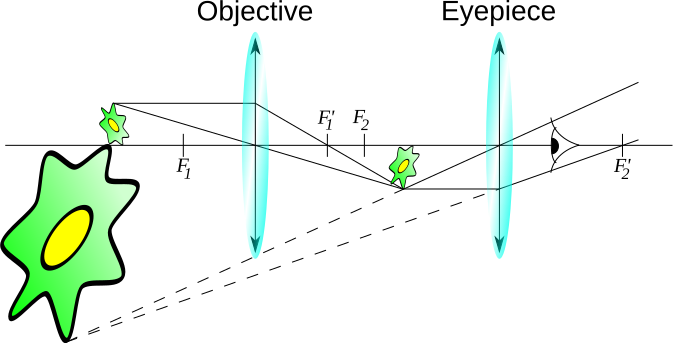
Diagram of a compound light microscope showing the optical path, eyepiece, objective lenses, condenser and illumination system used to magnify specimens. Source.
Magnification: The number of times larger an image appears compared with the real size of the object.
Resolution: The minimum distance at which two points can be distinguished as separate. Higher resolution produces clearer images.
Magnification and Resolution in Light Microscopy
Light microscopes typically achieve a maximum magnification of around ×1500 and a resolution of approximately 200 nm. These limitations arise because the wavelength of visible light restricts the amount of detail that can be resolved. Although resolution is limited, light microscopy enables:
Observation of whole cells and tissues
Viewing in colour
Real-time imaging of living processes, such as cell division or cytoplasmic streaming
A normal sentence must follow before moving on to electron microscopy to ensure logical flow between sections.
Electron Microscopy
Electron microscopes use beams of electrons instead of light, allowing much greater magnification and resolution because electrons have a far shorter wavelength than visible light. This enables visualisation of ultrastructure, including organelles such as mitochondria and ribosomes.
General Features of Electron Microscopes
Electron microscopes require specimens to be placed in a vacuum, meaning only dead material can be observed. Images are produced as either two-dimensional or three-dimensional representations of surface or internal structures.
Much higher resolution (up to 0.1 nm) compared with light microscopes
Black and white images (colour can be added artificially)
Complex specimen preparation involving fixation, dehydration and staining with heavy metal ions
Transmission Electron Microscope (TEM)
TEMs pass electrons through a thin specimen to produce a highly detailed image of internal structures. They are particularly useful for studying the arrangement of organelles and features such as cristae or thylakoids.
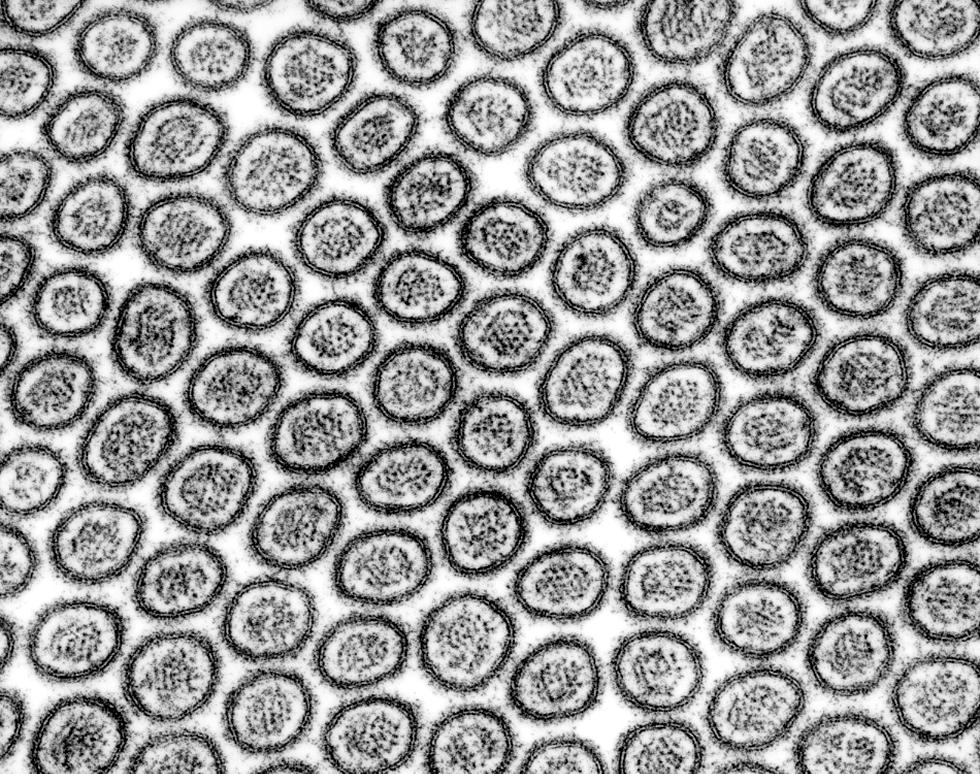
TEM micrograph showing cellular ultrastructure in thin section, revealing internal detail due to the high resolution achieved when electrons pass through the specimen. Source.
Key characteristics of TEM:
Very high resolution, allowing detailed internal cell imaging
Produces a 2D image
Suitable for examining cellular ultrastructure such as membranes, ribosomes and internal organelle detail
Scanning Electron Microscope (SEM)
SEMs scan a focused beam of electrons across the specimen surface, producing a detailed 3D image that reveals surface texture and architecture, such as cilia, microvilli or bacterial cell walls.
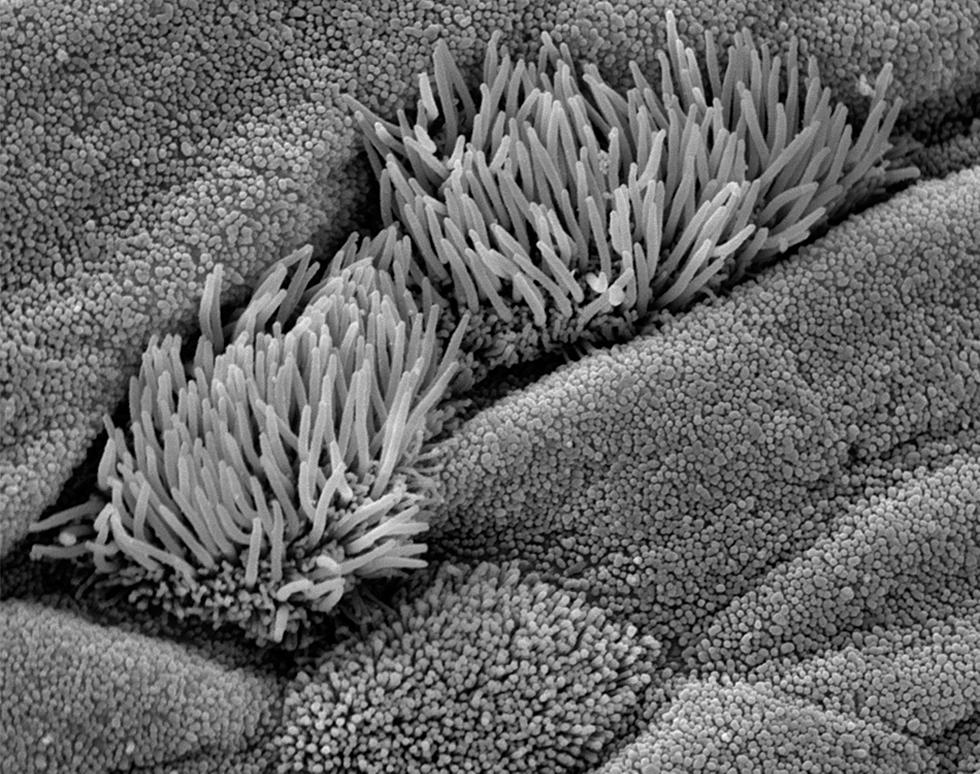
SEM micrograph showing surface features of epithelial cells, demonstrating the three-dimensional imaging capability of SEM for studying external morphology. Source.
Key characteristics of SEM:
Lower resolution than TEM, but still greater than light microscopy
Produces a 3D image of surfaces
Ideal for visualising external morphology
A sentence here highlights the relationship between TEM, SEM and their biological applications, reinforcing how they complement each other.
Comparing Light, TEM and SEM
Biologists select a microscope based on the structure of interest and the level of detail required. The most important differences relate to magnification, resolution, image type and whether the specimen must be dead.
Light microscope: lower magnification and resolution; views whole, living or dead cells; images in colour; internal detail is limited
TEM: highest resolution; 2D images of internal ultrastructure; only dead specimens; extremely detailed
SEM: high resolution; 3D surface images; only dead specimens; excellent for external features
Electron microscopes therefore reveal the complexity of cell ultrastructure far beyond the capability of light microscopy.
Magnification Formula
Students must be confident applying the standard magnification equation when interpreting images seen under any microscope.
EQUATION
—-----------------------------------------------------------------
Magnification (M) = Image size / Actual size
Image size = Measured size of the picture or photomicrograph
Actual size = Real size of the object
—-----------------------------------------------------------------
This relationship allows meaningful comparison between images from light, TEM and SEM instruments and links observed features to true scale.
Applications in Cell Observation
Light microscopes: ideal for cell division, tissues and living samples
TEM: essential for studying organelle structure and internal membranes
SEM: used for observing surfaces, including microbial cell walls and extracellular features
By selecting the appropriate microscope, biologists can match the instrument’s properties to their observational needs, ensuring maximum clarity and relevance in cell studies.
Practice Questions
Question 1 (2 marks)
State the difference between magnification and resolution in microscopy.
Question 1 (2 marks)
Award one mark for each correct point:
Magnification is how many times larger the image is compared to the actual object. (1)
Resolution is the ability to distinguish two close points as separate. (1)
Question 2 (5 marks)
Compare the use of a light microscope and a transmission electron microscope (TEM) when observing cells. In your answer refer to both the advantages and limitations of each instrument.
Question 2 (5 marks)
Award up to five marks from the following points:
Light microscopes use light, while TEMs use electrons. (1)
TEMs have higher resolution than light microscopes, allowing detailed ultrastructure to be seen. (1)
Light microscopes can view living specimens, but TEMs can only observe dead specimens due to the vacuum. (1)
TEMs produce 2D images of internal structures, whereas light microscopes can show whole cells but with limited detail. (1)
Light microscopy requires simpler preparation, whereas TEM sample preparation is complex and time-consuming. (1)
FAQ
Electron beams are easily scattered by particles in air, which would blur the image and drastically reduce resolution. Creating a vacuum ensures electrons travel in straight paths towards the specimen and detector.
Because living cells cannot survive in a vacuum, all specimens for TEM and SEM must be dead, dehydrated and stabilised before imaging.
Electron stains contain heavy metals such as lead or osmium. These atoms strongly scatter electrons, creating darker contrast in electron micrographs.
Unlike light stains, which add colour, electron stains do not produce colour images. Instead, they enhance differences in electron density to reveal ultrastructure more clearly.
SEM detects secondary electrons emitted from the specimen's surface. These electrons vary depending on surface contours, so raised areas emit more and appear brighter.
By scanning line by line and measuring surface electron output, the SEM reconstructs a three-dimensional image with shadows and depth.
TEM relies on electrons passing through the specimen. If the sample is too thick, electrons are absorbed or scattered, preventing a clear image.
Ultrathin sections, often less than 100 nm, allow electrons to transmit effectively, revealing internal membranes, organelles and fine structures.
Resolution is limited by the wavelength of visible light. Even with high-quality lenses, light cannot resolve objects smaller than roughly half its wavelength.
Because electrons have a far shorter wavelength, electron microscopes can exceed the resolution limit of light microscopy and reveal much finer detail.
