
OCR Specification focus:
‘Contrast prokaryotic and eukaryotic cells and interpret TEM/SEM images of cell components.’
Eukaryotic and prokaryotic cells share core features but differ greatly in internal organisation. Understanding these differences is essential for interpreting electron micrographs and recognising key structures in cells.
Cells are broadly divided into prokaryotes and eukaryotes, differing in size, organisation and internal membranes. Electron microscopy enables detailed study of ultrastructure for comparison and identification.
Key distinctions between prokaryotic and eukaryotic cells
Prokaryotes such as bacteria are simpler and smaller than eukaryotic cells. Eukaryotes include animals, plants, fungi and protoctists, and possess internal membranes and organelles.
Structural differences
Organelle: A specialised structure within a cell that performs a particular function.
Prokaryotes do not possess membrane-bound organelles, while eukaryotes contain many. Their major distinguishing features include the following:
Nucleus
Prokaryotes: No true nucleus; DNA is found as a single circular molecule in the nucleoid.
Eukaryotes: DNA contained within a double membrane-bound nucleus.
DNA organisation
Prokaryotes: Circular DNA, not associated with histone proteins; may have plasmids.
Eukaryotes: Linear chromosomes associated with histone proteins.
Ribosomes
Prokaryotes: 70S ribosomes in the cytoplasm.
Eukaryotes: 80S ribosomes in the cytoplasm and on rough ER.
Cell size
Prokaryotes: Typically 0.5–5 μm.
Eukaryotes: Typically 20–100 μm.
Organelles and membranes
Prokaryotes: No mitochondria, Golgi apparatus, ER or chloroplasts; mesosomes may be present in some species.
Eukaryotes: Mitochondria for aerobic respiration; plants and some protoctists contain chloroplasts.
Cell wall
Prokaryotes: Made of peptidoglycan.
Plants (eukaryotic): Made of cellulose; fungi have chitin; animals lack cell walls.
Reproduction
Prokaryotes: Divide by binary fission.
Eukaryotes: Divide by mitosis and meiosis.
Flagella
Prokaryotic flagella: Structurally simple, made of flagellin, rotating for movement.
Eukaryotic flagella: More complex with a 9+2 microtubule arrangement.
These structural differences allow rapid prokaryotic replication and metabolic flexibility, contrasting with the compartmentalisation and specialisation seen in eukaryotes.
Interpreting electron micrographs (TEM and SEM)
Electron microscopy is essential for visualising ultrastructure. Light microscopes lack the resolution to distinguish many organelles, so Transmission Electron Microscopy (TEM) and Scanning Electron Microscopy (SEM) are used in cell ultrastructure investigations.
Resolution: The ability to distinguish two points as separate in an image.
Transmission Electron Microscope (TEM)
TEM produces highly detailed 2D images of internal structures.
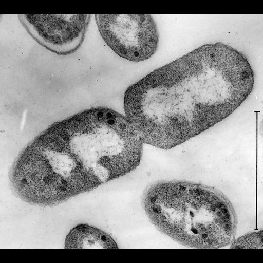
Transmission electron micrograph of a longitudinal thin section of a dividing E. coli cell, showing the dense nucleoid region, surrounding 70S ribosomes, and the cell wall near the boundary. Internal simplicity and absence of a nucleus distinguish prokaryotes in TEM images. Source.
Electrons pass through thin specimens, revealing organelles clearly. Students should recognise:
Nucleus with dark nucleolus
Mitochondria with folded cristae
Chloroplasts showing grana and stroma
Rough ER with ribosomes attached
70S vs 80S ribosomes, visible as smaller or larger dark dots
Bacterial cell walls and nucleoid regions in prokaryotes
Because TEM images are flat and sectional, organelles appear as slices in varying orientations.
Scanning Electron Microscope (SEM)
SEM provides 3D surface images by bouncing electrons off a specimen’s surface.
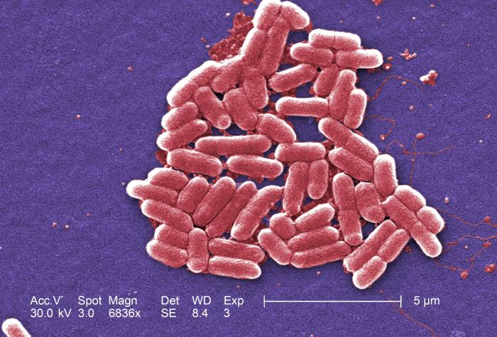
Scanning electron micrograph of rod-shaped E. coli, showing the three-dimensional contours of the bacterial surface. SEM highlights external morphology rather than internal detail, producing depth and texture absent in TEM. Source.
SEM is useful for observing:
Cell surfaces and microvilli
Prokaryotic shapes (cocci, bacilli, spirilla)
External features such as cilia or flagella
Although SEM shows less internal detail, it produces dramatic surface images that help identify overall morphology.
Recognising prokaryotes in micrographs
Prokaryotic cells in EM images typically show:
No visible nucleus
No membrane-bound organelles
70S ribosomes appearing small and scattered
Cell wall close to the outer boundary
Possible flagellum, capsule or plasmids
Their small size and simple internal appearance distinguish them clearly from eukaryotic cells.
Recognising eukaryotes in micrographs
Eukaryotic cells show:
A distinct nucleus with a nuclear envelope
Mitochondria or chloroplasts
80S ribosomes
Extensive endomembrane network (ER and Golgi apparatus)
Greater internal complexity and compartmentalisation
Using scale bars in EM images
EM images always include a scale bar to determine size. Magnification alone is insufficient, as images can be resized. Students must use the scale bar to make measurements and compare structures reliably when distinguishing cell types.
Summary of visual cues for rapid identification
Prokaryote: small, simple, no nucleus, no membrane-bound organelles, 70S ribosomes
Eukaryote: larger, complex, nucleus present, membrane-bound organelles, 80S ribosomes
Practice Questions
Question 1 (2 marks)
State two structural differences between prokaryotic and eukaryotic cells.
Question 1 (2 marks)
Award 1 mark for each correct difference, up to 2 marks:
Prokaryotes have no nucleus, whereas eukaryotes have a nucleus.
Prokaryotes have 70S ribosomes, whereas eukaryotes have 80S ribosomes.
Prokaryotic DNA is circular and not associated with histones, whereas eukaryotic DNA is linear and associated with histones.
Prokaryotes lack membrane-bound organelles, whereas eukaryotes possess them.
Prokaryotes have cell walls made of peptidoglycan, whereas eukaryotic cell walls (if present) are not made of peptidoglycan.
Question 2 (5 marks)
Fig. 1 shows a transmission electron micrograph (TEM) of a cell. Describe how features visible in a TEM image could be used to determine whether the cell is prokaryotic or eukaryotic. Include references to at least three identifiable structures in your answer.
Question 2 (5 marks)
Award marks for the following valid points, up to 5 marks:
Presence or absence of a nucleus can be observed; a nucleus indicates a eukaryotic cell. (1)
80S ribosomes in eukaryotes are larger than 70S ribosomes in prokaryotes. (1)
Presence of membrane-bound organelles such as mitochondria or chloroplasts identifies a eukaryote. (1)
A visible nucleoid region without a surrounding membrane suggests a prokaryote. (1)
Cell wall composition cannot be directly determined from TEM, but a thin, simple wall suggests a prokaryote, whereas a more complex structure suggests a eukaryote. (1)
Reference to TEM producing 2D internal detail showing internal ultrastructure. (1)
(max 5 marks)
FAQ
TEM images show thin internal sections with organelles clearly visible as darker or lighter regions. They appear flat and two-dimensional, showing internal ultrastructure such as cristae in mitochondria or membranes of the nucleus.
SEM images show a three-dimensional surface, often with shadows and contours. They do not reveal internal structures, instead highlighting the texture and shape of cells, such as the surface of bacterial cell walls or cilia.
Prokaryotes lack membrane-bound organelles, so their interiors look more uniform, with only ribosomes and a nucleoid region visible.
They also have a smaller volume, leading to fewer distinguishable internal features. In contrast, eukaryotes contain numerous organelles, membranes, and compartmentalised regions that are easily identified in EM images.
Electron micrographs can reveal a relatively thick, dense boundary immediately inside the capsule or membrane.
Some SEM images show surface texture differences between Gram-positive and Gram-negative bacteria:
Gram-positive: smoother, thicker wall appearance
Gram-negative: thinner wall with an uneven surface due to the outer membrane
These observations can support identification, although staining information is still required for certainty.
Ribosomes are made of RNA and protein, which strongly scatter electrons. This creates dark, granular dots even at high magnification.
Their density and size also help identification:
Small and numerous in prokaryotes (70S)
Larger and sometimes attached to rough ER in eukaryotes (80S)
Students often mistake vesicles for nuclei or confuse mitochondria with chloroplasts due to similar shapes.
Other frequent errors include:
Assuming any dark region is a nucleolus
Expecting SEM to show organelles
Ignoring scale bars, leading to incorrect assumptions about cell type
Careful attention to membrane boundaries, internal detail, and scale improves accuracy.
