
Edexcel Syllabus focus:
'Understand how the structures of arteries, veins and capillaries are related to their different functions in circulation.'
Blood vessels are not built the same way. Their wall thickness, lumen size, elasticity, and permeability all match the pressure of blood flow and the need for exchange with tissues.
Blood vessels in circulation
In a mammalian circulatory system, blood must move efficiently from the heart to tissues and then return again. Different blood vessels are specialized for different parts of this pathway:
Arteries carry blood away from the heart under high pressure.
Veins return blood to the heart under lower pressure.
Capillaries are the sites of exchange between blood and body cells.
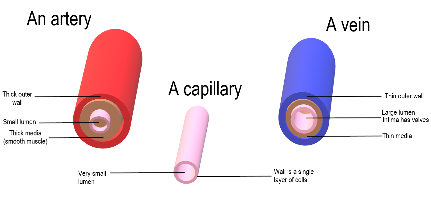
Cross-sections comparing an artery, a vein, and a capillary, highlighting the relative wall thickness and lumen size. This supports the idea that arteries are built to withstand high pressure, veins are adapted for low-pressure return, and capillaries are specialized for exchange with a very thin wall. Source
The key idea is that each vessel has a structure that suits its function. Wall composition, lumen diameter, and the presence or absence of valves are all important.
Arteries
Structure of arteries
Arteries have thick walls compared with their lumen. The wall contains:
a thick layer of smooth muscle
many elastic fibers
a tough outer layer containing collagen
a smooth inner lining called the endothelium
They usually have a small lumen relative to wall thickness, and they do not contain valves along most of their length.
How artery structure matches function
Blood leaving the heart is pumped at high pressure, so arteries must be strong enough to resist bursting. Their thick wall and collagen give this strength.
The high amount of elastic tissue is important because artery walls stretch when blood is forced into them during ventricular contraction and then recoil as the pressure falls. This recoil helps maintain blood pressure and keeps blood moving between heartbeats, giving a smoother flow rather than a stop-start movement.
Smooth muscle in the artery wall allows the vessel to constrict or dilate. Changing the diameter of an artery helps control blood flow to different tissues.
A relatively narrow lumen helps maintain the high pressure of blood. The smooth endothelium reduces friction, so blood can flow more easily.
Veins
Structure of veins
Veins also have a lumen and an endothelial lining, but their walls are very different from those of arteries:
the wall is much thinner
there is less smooth muscle and less elastic tissue
the lumen is wide
many veins contain valves
Because blood pressure in veins is low, veins often appear less circular than arteries and may be flattened if empty.
How vein structure matches function
By the time blood reaches veins, much of the original pressure produced by the heart has been lost. Veins therefore do not need thick muscular, elastic walls. Their thinner walls are sufficient for low-pressure flow.
The wide lumen reduces resistance to blood flow, helping blood return to the heart even though the pressure is low. This is especially important over long distances, such as from the legs.
Vein valves are crucial. They prevent backflow of blood and ensure that blood moves in one direction, especially when it is moving against gravity.
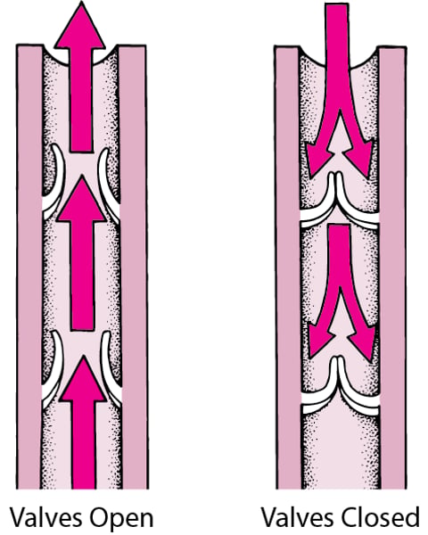
Diagram of one-way venous valves showing forward flow opening the valve cusps and reverse flow closing them. This visualizes how valves maintain unidirectional blood movement in low-pressure veins, particularly in the limbs where gravity promotes backflow. Source
This is most important in the limbs. Contraction of surrounding skeletal muscles squeezes veins, and the valves make sure this squeezing pushes blood toward the heart rather than backward.
Since veins have less elastic tissue, they do not show the same stretch-and-recoil behavior as arteries. Their structure is suited to returning blood slowly and steadily at low pressure.
Capillaries
Structure of capillaries
Capillaries are microscopic vessels that form dense networks called capillary beds within tissues. Their structure is very simple:
the wall is one cell thick, made only of endothelial cells
they have an extremely narrow lumen
they are highly branched, giving a very large total surface area
small gaps between endothelial cells make the wall relatively permeable
Red blood cells often pass through capillaries in single file because the lumen is so narrow.
How capillary structure matches function
Capillaries are the main sites where substances move between blood and tissues. Their very thin wall creates a short diffusion distance, so materials such as oxygen and glucose can move out of the blood efficiently, while carbon dioxide can move into the blood.
The narrow lumen slows blood flow and keeps red blood cells close to the wall. This improves the rate of exchange because substances do not have far to travel.
The large number of capillaries in a tissue creates a large surface area for exchange. A bigger surface area allows more movement of substances per unit time.
Their permeable walls allow plasma and dissolved substances to leave the blood and form tissue fluid. Useful materials can then diffuse to cells, while waste products diffuse back into the blood.
Capillaries are not built to withstand high pressure like arteries. Instead, they are built for efficient exchange.
Comparing vessel structure and function
The easiest way to understand these vessels is to link each major structural feature to its function:
Thick, elastic, muscular wall -> artery -> withstands and maintains high pressure
Thin wall, wide lumen, valves -> vein -> returns blood at low pressure and prevents backflow
One-cell-thick wall, narrow lumen, extensive branching -> capillary -> allows rapid exchange between blood and tissues
A common exam approach is to compare matching features directly:
more elastic tissue in arteries than veins because arteries experience pressure surges
valves in veins but not arteries because venous blood is more likely to flow backward
a one-cell-thick wall in capillaries, unlike arteries and veins, because capillaries are specialized for exchange rather than transport over distance
Practice Questions
State two structural features of capillaries that make them efficient exchange surfaces. (2 marks)
Wall is one cell thick / made of a single layer of endothelial cells (1)
Narrow lumen / red blood cells pass close to the wall / blood flow is slowed (1)
Large network / highly branched / large total surface area (1)
Permeable wall / gaps between endothelial cells (1)
Max 2 marks
Explain how the structures of arteries, veins, and capillaries are related to their different functions in circulation. (6 marks)
Award 1 mark for each valid linked point, up to 6 marks:
arteries have thick walls / collagen to withstand high pressure
arteries contain elastic tissue to stretch and recoil
recoil in arteries helps maintain pressure / smooth blood flow
arteries contain smooth muscle to change diameter / control blood flow
arteries have a relatively narrow lumen to help maintain pressure
veins have thinner walls because blood is at lower pressure
veins have less elastic tissue and less smooth muscle than arteries
veins have a wide lumen to reduce resistance to flow
veins have valves to prevent backflow
capillaries have walls that are one cell thick, giving a short diffusion distance
capillaries have a narrow lumen, keeping blood close to the wall / slowing flow
capillaries form extensive networks, giving a large surface area for exchange
capillary walls are permeable, allowing movement of substances between blood and tissues
FAQ
A pulse is a pressure wave produced when the ventricles contract and force blood into the arteries.
Arteries can stretch and then recoil because they contain a lot of elastic tissue. This makes the pressure wave detectable as a pulse. By the time blood has passed through capillaries and entered veins, the pressure is much lower and the pulse has been lost.
Veins are usually preferred because they are safer and easier to access.
they are closer to the body surface
blood is under lower pressure
bleeding is easier to stop after the needle is removed
there is usually less pain and lower risk of serious damage
Arteries are deeper, have a stronger pulse, and bleed more heavily if punctured.
Veins in the legs must return blood to the heart against gravity.
If vein valves become weak or damaged, blood can fall backward and collect in the vein. This increases pressure inside the vein, causing it to widen and become twisted. Long periods of standing, aging, and pregnancy can all increase the chance of this happening.
Arteries have thicker walls with more smooth muscle, elastic tissue, and collagen. This gives them more structural support, so they tend to stay round even when not full.
Veins have thinner walls and less elastic support. When there is little blood inside them, they are more likely to flatten or collapse.
Capillaries have very thin, permeable walls, so they are easily damaged by knocks or inflammation.
If capillaries break:
red blood cells can leak into surrounding tissues, causing a bruise
plasma can leave the blood, increasing tissue fluid and causing swelling
This is one reason capillary beds are efficient for exchange but also more delicate than arteries and veins.