
Edexcel Syllabus focus:
'Relate the structure and operation of the mammalian heart, including the major blood vessels, to its function.'
The mammalian heart is a muscular double pump. Its chambers, valves, and linked blood vessels are arranged so blood moves rapidly, efficiently, and in one direction to the lungs and body.
The heart as a double pump
The mammalian heart acts as two pumps working side by side. The right side sends deoxygenated blood to the lungs for gas exchange, while the left side sends oxygenated blood to the rest of the body. This arrangement supports a high rate of respiration because blood can be delivered quickly and at appropriate pressure to different parts of the circulation.
Double circulation: A transport system in which blood passes through the heart twice during one complete circuit of the body.
Keeping the pulmonary and systemic circuits separate makes transport more efficient than mixing blood in a single pump.
Chambers of the mammalian heart
Humans and other mammals have a four-chambered heart. The two atria are the upper chambers and mainly receive blood. The two ventricles are the lower chambers and pump blood out of the heart.
The right atrium receives blood from the vena cava. The left atrium receives blood from the pulmonary veins. The right ventricle pumps blood into the pulmonary artery, and the left ventricle pumps blood into the aorta.
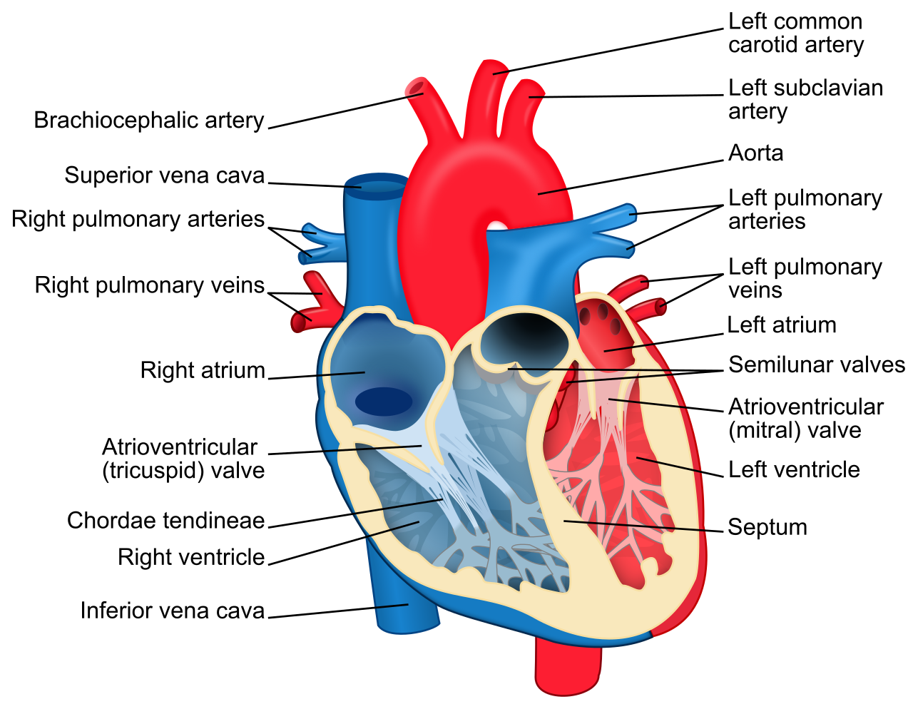
Labeled cross-section of the heart showing the four chambers, septum, major valves, and the great vessels entering/leaving the heart. The diagram uses contrasting colors to distinguish the deoxygenated (right heart → lungs) and oxygenated (left heart → body) pathways, reinforcing the idea of double circulation. Source
A muscular wall called the septum separates the left and right sides. This prevents oxygenated and deoxygenated blood from mixing, so tissues receive blood with the highest possible oxygen concentration.
The atria have relatively thin walls because they only need to move blood a short distance into the ventricles.
Major blood vessels
Four major vessels are closely linked to heart function:
Vena cava – returns deoxygenated blood from the body to the right atrium.
Pulmonary artery – carries deoxygenated blood from the right ventricle to the lungs.
Pulmonary veins – carry oxygenated blood from the lungs to the left atrium.
Aorta – carries oxygenated blood from the left ventricle to the body.
These vessels are positioned so blood enters the atria and leaves from the ventricles, matching the receiving and pumping roles of the chambers.
Valves and one-way flow
Efficient circulation depends on one-way blood flow. The heart contains valves, which prevent blood from moving backward when pressure changes during contraction and relaxation.
Valve: A flap-like structure that opens and closes in response to pressure differences, preventing backflow of blood.
There are two main sets of valves:
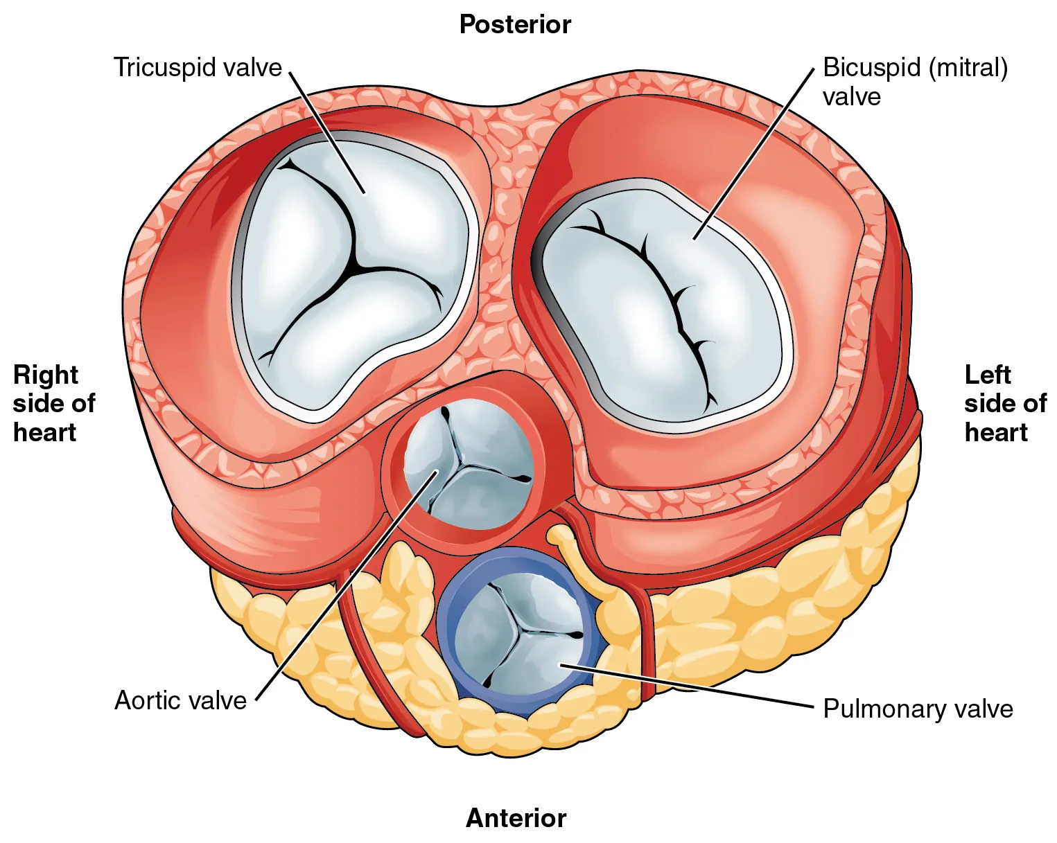
Diagram focusing on the four heart valves, labeling the atrioventricular (tricuspid and mitral/bicuspid) and semilunar (pulmonary and aortic) valves. It helps visualize how valve placement enforces one-way flow between atria → ventricles and ventricles → great arteries. Source
Atrioventricular (AV) valves between each atrium and ventricle. The right AV valve is the tricuspid valve and the left AV valve is the bicuspid or mitral valve.
Semilunar valves at the bases of the pulmonary artery and aorta.
When ventricular pressure rises, the AV valves close and the semilunar valves open. When ventricular pressure falls, the semilunar valves close, preventing backflow from the arteries.
The AV valves are supported by tendinous cords attached to papillary muscles. These stop the valves from turning inside out when ventricles contract.
If valves did not work properly, some blood would flow backward, reducing the efficiency of transport.
Ventricular wall thickness and pressure
The left ventricle has a much thicker muscular wall than the right ventricle. It must generate high pressure to push blood through the entire systemic circulation.
The right ventricle only pumps blood to the nearby lungs, so a thinner wall is sufficient. Lower pressure in the pulmonary circulation helps protect delicate lung capillaries and supports efficient gas exchange.
This difference in wall thickness is a clear example of structure matching function.
The left atrium and right atrium both have thinner walls than the ventricles because they pump over much shorter distances.
Coronary circulation and heart muscle
The heart wall is made mostly of cardiac muscle, also called the myocardium. Like all active tissues, it needs a continuous supply of oxygen and glucose for aerobic respiration.
The blood inside the chambers cannot supply the whole heart wall fast enough by diffusion alone, so the heart has its own blood supply.
Coronary arteries branch from the base of the aorta and deliver oxygenated blood to the heart muscle.
Coronary veins return deoxygenated blood from the heart muscle to the right side of the heart.
This dedicated circulation allows the heart to keep contracting throughout life.
Path of blood through the heart
The route of blood through the heart is:
body → vena cava → right atrium
right atrium → tricuspid valve → right ventricle
right ventricle → pulmonary semilunar valve → pulmonary artery → lungs
lungs → pulmonary veins → left atrium
left atrium → bicuspid valve → left ventricle
left ventricle → aortic semilunar valve → aorta → body
Because the chambers and valves are arranged in this sequence, blood moves efficiently to the lungs for oxygenation and then to the body for delivery of oxygen and other transported substances.
Coordinated contraction of atria followed by ventricles keeps this flow effective.
Practice Questions
Explain why the wall of the left ventricle is thicker than the wall of the right ventricle. (2 marks)
Left ventricle pumps blood to the whole body / systemic circulation. (1)
It must generate higher pressure / more force than the right ventricle, which only pumps blood to the lungs. (1)
Describe how the structure of the mammalian heart and its major blood vessels help maintain efficient one-way transport of blood. (5 marks)
Heart has four chambers; atria receive blood and ventricles pump blood out. (1)
Septum separates left and right sides / prevents mixing of oxygenated and deoxygenated blood. (1)
Correct role of major vessels, for example vena cava to right atrium, pulmonary artery to lungs, pulmonary veins to left atrium, or aorta to body. (1)
AV valves and semilunar valves prevent backflow / maintain one-way movement of blood. (1)
Left ventricle has a thicker wall to generate high pressure for systemic circulation; right ventricle has a thinner wall for lower pressure to the lungs. (1)
FAQ
Arteries and veins are named by the direction of blood flow relative to the heart, not by oxygen content.
An artery carries blood away from the heart, so the pulmonary artery is still an artery even though it carries deoxygenated blood. A vein carries blood toward the heart, so the pulmonary vein is still a vein even though it carries oxygenated blood.
Each lung is commonly drained by two main pulmonary veins: a superior and an inferior vein.
That gives a total of four pulmonary veins entering the left atrium in most people. Small anatomical variations can occur, but four is the usual pattern described in human anatomy.
Semilunar valves are shaped like small pockets. When blood starts to flow backward, the pockets fill and press tightly together.
Because of this design, they do not risk flipping back into the ventricles in the same way AV valves would. AV valves face a different pressure direction during ventricular contraction, so they need tendinous cords for support.
The fibrous skeleton is a framework of dense connective tissue around the heart valves and between the atria and ventricles.
It helps by:
anchoring valve structures
providing support for cardiac muscle
electrically insulating the atria from the ventricles except through the normal conduction pathway
This helps keep contraction organized.
The apex is the pointed lower tip of the heart, and it is formed mainly by the left ventricle because that chamber has the greatest muscle mass.
Its thicker wall and more forceful pumping role make it the most prominent lower chamber. This is why the heartbeat is often felt most strongly near the apex region.