
OCR Specification focus:
‘Identify chambers, vessels and valve positions; practise dissection, examination and accurate biological drawing of the heart’s external and internal structures.’
The mammalian heart is a muscular organ responsible for maintaining blood circulation through double circulation, ensuring oxygenated and deoxygenated blood remain separate. Its anatomy demonstrates remarkable specialisation for efficiency and control.
External Anatomy of the Mammalian Heart
Overall Structure
The mammalian heart is a four-chambered organ located in the thoracic cavity between the lungs, enclosed within a pericardium — a double-layered sac providing protection and lubrication. The heart’s orientation is slightly tilted, with the apex pointing to the left.
Pericardium: A double-layered membrane enclosing the heart, reducing friction during contraction and maintaining its position within the thorax.
The heart’s exterior shows two atria (upper chambers) and two ventricles (lower chambers). The coronary arteries and veins are visible on the surface, supplying oxygen and nutrients to cardiac tissue. Blockage of these vessels can cause a myocardial infarction (heart attack) due to oxygen deprivation in cardiac muscle.
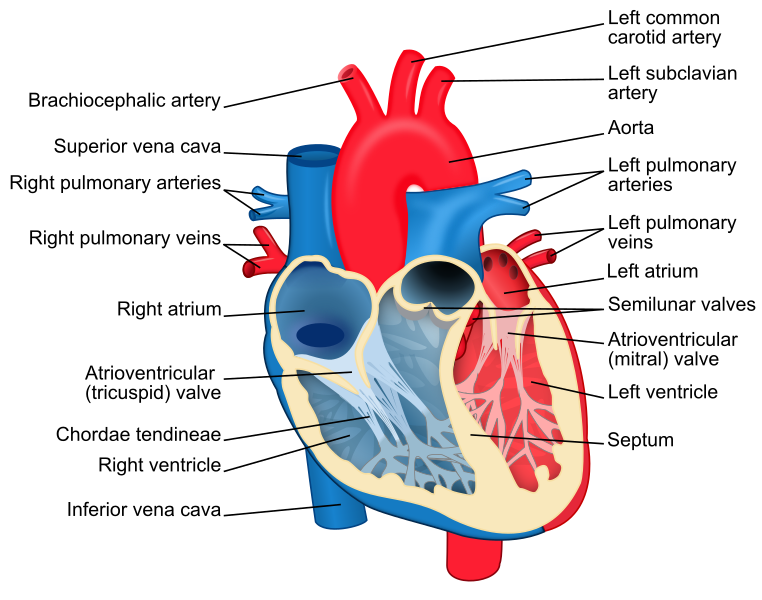
A labelled diagram of the human heart showing the right and left atria and ventricles, major vessels (aorta, pulmonary artery/veins, vena cava), and valves. Colours distinguish oxygenated and deoxygenated pathways, reinforcing double circulation. Clean labelling supports accurate biological drawing and revision. Source.
Major External Blood Vessels
The heart connects to four main vessels visible externally:
Aorta – carries oxygenated blood from the left ventricle to the body.
Pulmonary artery – transports deoxygenated blood from the right ventricle to the lungs.
Pulmonary veins – bring oxygenated blood from the lungs to the left atrium.
Vena cava (superior and inferior) – return deoxygenated blood from the body to the right atrium.
These vessels ensure unidirectional blood flow and separation of systemic and pulmonary circuits.
The Coronary Circulation
The right and left coronary arteries branch from the aorta to supply oxygenated blood to the myocardium. Venous return occurs via coronary veins, which drain into the coronary sinus, emptying into the right atrium. This ensures that the heart muscle itself receives an uninterrupted oxygen supply despite constant contraction.
Internal Anatomy of the Mammalian Heart
Chambers and Their Features
The heart is divided into left and right sides by a septum, preventing the mixing of oxygenated and deoxygenated blood.
Right atrium: Receives deoxygenated blood from the vena cava.
Right ventricle: Pumps this blood to the lungs via the pulmonary artery.
Left atrium: Receives oxygenated blood from the pulmonary veins.
Left ventricle: Pumps oxygenated blood to the body through the aorta.
The left ventricle has a thicker muscular wall than the right to generate higher pressure needed for systemic circulation, while the right ventricle pumps blood at lower pressure to protect the delicate lung capillaries.
Septum: The muscular wall separating the left and right sides of the heart, preventing mixing of oxygenated and deoxygenated blood.
Valves and Direction of Flow
Valves within the heart maintain unidirectional flow, preventing backflow during contraction and relaxation phases:
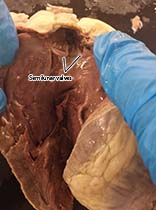
A close view of the semilunar valves at an arterial outflow, illustrating the three cusps that close to prevent backflow into the ventricle. This supports locating the aortic and pulmonary valve positions during dissection. Labels focus on the valve cusps; any additional background tissue visible is incidental and not beyond syllabus scope. Source.
Atrioventricular (AV) valves:
Tricuspid valve – between right atrium and ventricle (three flaps).
Bicuspid (mitral) valve – between left atrium and ventricle (two flaps).
Semilunar valves:
Pulmonary valve – between right ventricle and pulmonary artery.
Aortic valve – between left ventricle and aorta.
Atrioventricular valves: Valves between atria and ventricles preventing blood from flowing back into the atria during ventricular contraction.
Tendinous cords attach valve flaps to papillary muscles within the ventricles, preventing inversion of the valves during high-pressure contractions.
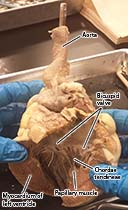
Opened left ventricle displaying bicuspid (mitral) valve cusps with chordae tendineae extending to papillary muscles, and the ridged trabeculae carneae of the ventricular wall. This real dissection view reinforces recognition of internal landmarks for drawing and identification. Labels are minimal and clearly placed; no extra systems are shown beyond syllabus needs. Source.
Cardiac Tissue Structure
The Myocardium
The myocardium consists of cardiac muscle tissue, a specialised involuntary muscle with intercalated discs allowing rapid transmission of electrical impulses and coordinated contraction. Unlike skeletal muscle, it is myogenic, meaning contractions originate within the heart itself rather than requiring neural stimulation.
Myogenic: Describes muscle that contracts without direct nervous stimulation, initiated instead by pacemaker activity within the heart.
This property ensures consistent and rhythmic contractions essential for effective pumping.
The Endocardium and Epicardium
The heart wall consists of three distinct layers:
Endocardium – smooth epithelial lining reducing friction and preventing blood clot formation.
Myocardium – thick middle layer responsible for contraction.
Epicardium – outer connective tissue layer forming part of the pericardium.
These layers work together to optimise mechanical strength, electrical conduction, and protection.
Functional Relationships Between Structures
Double Circulation Pathway
The mammalian heart supports double circulation, comprising:
Pulmonary circuit:
Right ventricle → Pulmonary artery → Lungs → Pulmonary veins → Left atrium
Ensures oxygenation of blood.
Systemic circuit:
Left ventricle → Aorta → Body → Vena cava → Right atrium
Delivers oxygen and nutrients to tissues.
This arrangement maintains high blood pressure in systemic circulation while protecting pulmonary capillaries from damage by keeping pulmonary pressure lower.
Double circulation: The separation of blood flow into pulmonary and systemic circuits, ensuring blood passes through the heart twice per complete cycle.
Adaptations for Efficiency
The heart demonstrates several key adaptations for efficiency:
Thicker left ventricle wall generates high pressure for systemic flow.
Valves ensure one-way flow, preventing inefficiency.
Coronary circulation supplies the heart muscle itself.
Septum maintains separation of oxygenated and deoxygenated blood.
Elastic and muscular tissue allow expansion and contraction without damage.
Dissection and Observation
When performing a heart dissection, students should:
Identify external features such as the coronary vessels and major arteries.
Make careful incisions to expose internal chambers and valves.
Observe thickness differences between ventricles and valve structures.
Use a hand lens or microscope to view the smooth endocardium and tendinous cords.
Drawing should include accurate proportions, labelled structures, and clear differentiation between left and right sides.
Practice Questions
Question 1 (2 marks)
Describe the role of the tendinous cords in the mammalian heart.
Mark Scheme:
1 mark for stating that tendinous cords prevent the atrioventricular valves from inverting or turning inside out during ventricular contraction.
1 mark for stating that they hold the valve flaps in position, ensuring unidirectional blood flow from atria to ventricles.
Question 2 (5 marks)
Explain how the structure of the left ventricle is related to its function.
Mark Scheme:
Award up to 5 marks from the following points:
The left ventricle has a thicker muscular wall than the right ventricle (1 mark).
This allows the generation of higher pressure needed to pump blood around the systemic circulation (1 mark).
The high pressure ensures oxygenated blood reaches all body tissues efficiently (1 mark).
The thick myocardium provides strong contractions without fatigue, maintaining consistent blood flow (1 mark).
The presence of valves prevents backflow of blood into the atrium during contraction, maintaining unidirectional flow (1 mark).
FAQ
The atria act as low-pressure receiving chambers for returning blood, while the ventricles function as high-pressure pumping chambers.
The right atrium collects deoxygenated blood from the body, whereas the left atrium receives oxygenated blood from the lungs.
The right ventricle pumps blood to the lungs at low pressure; the left ventricle generates higher pressure to drive systemic circulation.
This structural difference ensures efficient movement of blood through both circuits without damaging delicate capillaries in the lungs.
The left ventricle must create much higher pressure to pump blood throughout the entire body, compared to the right ventricle, which only pumps to the lungs.
Its thick myocardial wall allows stronger contractions.
The right ventricle’s thinner wall prevents excessive pressure in the pulmonary circuit, avoiding capillary damage.
This asymmetry is vital to maintain effective double circulation.
The coronary arteries supply oxygenated blood directly to the heart muscle (myocardium) itself. Unlike systemic arteries, their role is purely to sustain cardiac tissue.
They branch from the aorta, just above the aortic valve.
Blockages can cause coronary heart disease or myocardial infarction, as the heart cannot store oxygen.
Their narrow lumen makes them particularly vulnerable to atherosclerosis.
Efficient coronary circulation is essential for continuous cardiac function.
Cardiac muscle is uniquely adapted for endurance and coordination:
Intercalated discs join cells, allowing rapid electrical transmission.
It is myogenic, so contractions originate within the heart itself.
Cells contain many mitochondria, supporting high aerobic activity.
The branching fibres enable strong, synchronised contractions.
These features allow the heart to beat rhythmically without fatigue.
Valve operation depends entirely on pressure differences across them rather than active control.
When pressure behind a valve exceeds pressure in front, the valve opens.
When pressure is higher ahead, it closes, preventing backflow.
This passive mechanism ensures continuous, unidirectional blood flow without energy expenditure.
It is an efficient system that coordinates automatically with each heartbeat.
