

CIE Syllabus focus:
'Understand how the m/z value of the molecular ion, M+, in a mass spectrum can be used to determine the relative molecular mass of a molecule.'
Mass spectrometry can reveal the mass of an entire molecule, not just its fragments. For this topic, the essential skill is identifying the molecular ion peak and using its value correctly.
The molecular ion
In a mass spectrometer, molecules are ionized so that positive ions can be detected. If a molecule loses one electron but does not break apart, it forms the molecular ion. This ion represents the whole molecule, so it is the peak that gives information about the mass of the complete molecule rather than the mass of a fragment.
Molecular ion: The positive ion formed when a molecule loses one electron without breaking apart.
The molecular ion is often written as M+. Because only an electron has been removed, the mass of the ion is almost the same as the mass of the original molecule. The lost electron has such a small mass that, at this level, it is ignored when interpreting the spectrum.
value: The ratio of the mass of an ion to its charge.
Mass spectrometers detect ions according to their values. For the molecular ion in typical A-Level questions, the charge is usually , so the value is numerically the same as the mass of that ion.
= mass of the ion
= charge of the ion
= relative molecular mass when the molecular ion has charge
This is why the molecular ion is so useful.
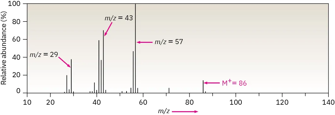
Mass spectrum of hexane with key peaks annotated, including the molecular ion at and prominent fragment ions at lower . The plot reinforces that, for a singly charged molecular ion, the molecular ion’s value equals the molecule’s relative molecular mass. Source
Once the correct M+ peak has been identified, its value can be used directly to find the molecule's relative molecular mass.
Using the M+ peak to find relative molecular mass
The relative molecular mass tells you how heavy a molecule is on the carbon-12 scale. In a mass spectrum, the M+ peak is the key signal because it comes from the unfragmented molecule. For a singly charged molecular ion, the numerical value of is the same as the value of .
Relative molecular mass, : The mass of a molecule compared with of the mass of a carbon-12 atom.
When reading a spectrum, do not assume that the tallest peak is the molecular ion.
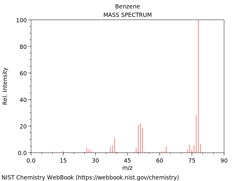
Electron-impact mass spectrum of benzene from the NIST Chemistry WebBook, showing relative intensity versus . The molecular ion peak for benzene appears at high (near 78), illustrating how for is used to infer molecular mass while other peaks correspond to fragments.
The tallest peak is the base peak, but it may be a fragment ion. Instead, focus on the peak that corresponds to the whole molecule and is usually found at a relatively high value.
How to identify the correct peak
Look for a peak from the unfragmented molecule.
Expect the M+ peak to appear at a higher than fragment peaks.
Remember that fragment ions come from the molecule breaking apart, so they have smaller masses.
Be aware that the molecular ion peak may be small if the molecule fragments easily.
If several nearby peaks appear in the molecular ion region, think about isotopes before deciding which peak represents the main molecular ion.
A correct interpretation depends on linking the peak to the whole molecule, not just choosing the highest or tallest signal. Once the M+ peak has been identified, its value gives the relative molecular mass directly in the common case where the charge is .
Why fragment peaks do not give relative molecular mass
Many peaks in a mass spectrum come from fragment ions.
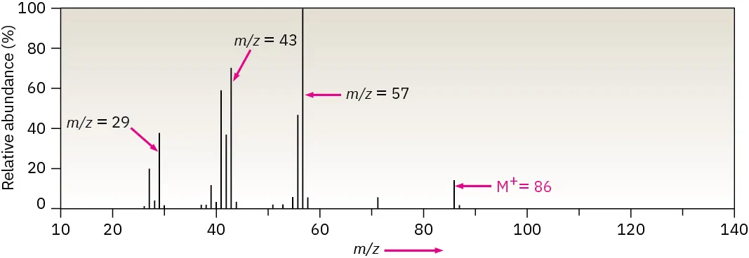
Fragmentation map for hexane showing formation of the molecular ion ( at ) and several common fragment ions at lower . The diagram emphasizes that the base peak can be a fragment (here at ) and therefore should not be used to determine . Source
These are formed when the molecular ion breaks into smaller positive ions. Fragment peaks are valuable because they can provide structural clues, but they do not represent the complete molecule. Using a fragment peak to find would therefore give a value that is too low.
This distinction is central to exam questions on mass spectrometry. You must identify whether a peak comes from the whole molecule or from part of it. Only the molecular ion peak should be used to determine relative molecular mass.
Common mistakes to avoid
Confusing the base peak with the molecular ion peak.
Choosing the highest-intensity peak instead of the correct M+ peak.
Using a fragment ion peak to calculate .
Forgetting that depends on charge as well as mass.
What the notation M+ tells you
The notation M+ is a reminder that the ion comes from the original molecule, represented by M, and carries a single positive charge. In most introductory mass spectra used at this level, this single charge is why the molecular ion peak can be read so directly. If the ion has charge , then the number shown on the spectrum is effectively the relative molecular mass of the molecule.
This makes the molecular ion peak one of the fastest ways to obtain basic information about an unknown molecular substance. Before using any value, however, always check that the peak really is the molecular ion and not a fragment or a nearby isotopic peak.
Practice Questions
A mass spectrum of a compound shows a molecular ion peak at .
State the relative molecular mass of the compound and explain your answer.
[2 marks]
Relative molecular mass is (1)
The molecular ion is the whole molecule / M+ ion, and for a singly charged ion (1)
A compound gives major peaks at , , , and in its mass spectrum.
(a) Identify the peak that should be used to determine the relative molecular mass of the compound.
(b) State the relative molecular mass of the compound.
(c) Explain why the other peaks cannot be used to determine the relative molecular mass.
[5 marks]
(a)
Peak at / the M+ peak (1)
(b)
(1)
(c)
The other peaks are fragment ions / ions from parts of the molecule (1)
Fragment ions have lower masses than the whole molecule (1)
Only the molecular ion represents the unfragmented molecule, so its value is used to find (1)
FAQ
These peaks are usually caused by isotopes in the molecule.
For example, a small M+1 peak often appears because some carbon atoms are $^{13}C$ instead of $^{12}C$. Molecules containing chlorine or bromine can show clear M+2 peaks because those elements have common heavier isotopes.
Yes. If an ion has charge $+2$, its $m/z$ value is half its mass.
At this level, the molecular ion is usually treated as singly charged, which is why the M+ peak can be used directly for $M_r$. In more advanced spectra, multiply charged ions can appear and must be identified carefully.
M+ forms when a molecule loses an electron. $[M+H]^+$ forms when a molecule gains a proton.
This matters because $[M+H]^+$ is one mass unit higher than the neutral molecule. So, in spectra that show $[M+H]^+$ instead of M+, the displayed peak is not read in exactly the same way.
A strong molecular ion peak suggests that the ion is relatively stable after ionization.
A very weak molecular ion peak suggests the ion fragments easily. That does not stop you from finding $M_r$, but it can make the molecular ion harder to spot among the fragment peaks.
Two different molecules can have the same whole-number relative molecular mass but slightly different exact masses.
High-resolution mass spectrometry measures those tiny differences very precisely. That allows chemists to distinguish between possible molecular formulas even when ordinary mass spectra show the same molecular ion value.