
OCR Specification focus:
‘Use adrenal glands to exemplify endocrine glands: outline cortex and medulla hormones and their functions.’
The adrenal glands are small, triangular endocrine glands located on top of the kidneys. They secrete vital hormones that regulate stress responses, metabolism, and blood pressure in humans.
Adrenal Gland Structure
Location and Overview
Each adrenal gland sits superior to a kidney and is enclosed in a fibrous capsule surrounded by adipose tissue. The gland has two distinct functional regions:
The adrenal cortex (outer layer)
The adrenal medulla (inner core)
These regions differ in both structure and the type of hormones they produce, reflecting their separate embryological origins — the cortex from the mesoderm, and the medulla from the ectoderm of the developing embryo.
The adrenal gland consists of an outer cortex with three zones—zona glomerulosa, zona fasciculata and zona reticularis—and an inner medulla.
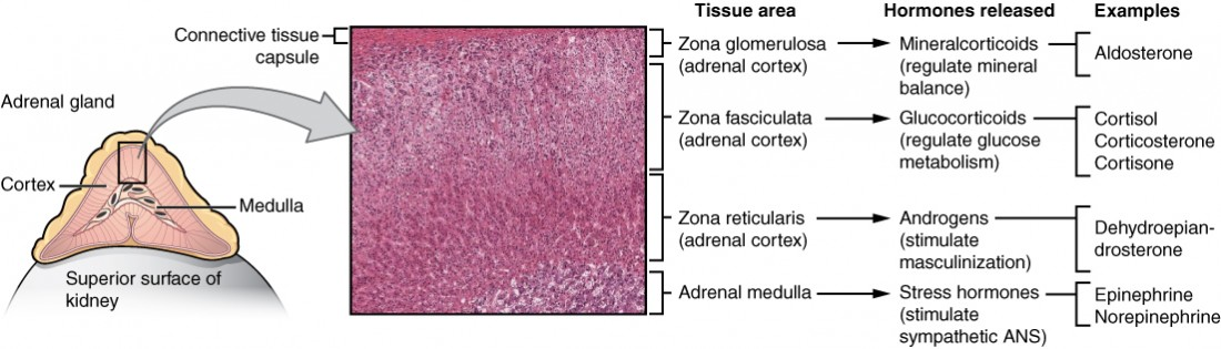
Labeled diagram and histology showing adrenal zonation and matching hormone outputs: mineralocorticoids (glomerulosa), glucocorticoids (fasciculata), androgens (reticularis), and medullary catecholamines. The concise arrows to hormone classes align with the OCR requirement to outline cortical versus medullary products. The micrograph in the centre provides real tissue context without excess detail. Source.
Adrenal Cortex
The adrenal cortex produces steroid hormones derived from cholesterol. It has three distinct zones, each secreting specific hormones.
1. Zona glomerulosa
This outermost layer produces mineralocorticoids, primarily aldosterone, which regulate sodium and potassium balance.
Aldosterone acts on the distal convoluted tubules and collecting ducts of nephrons, stimulating sodium reabsorption and potassium excretion.
This helps maintain blood volume and blood pressure, as sodium reabsorption drives water reabsorption by osmosis.
Mineralocorticoids: Steroid hormones that regulate electrolyte and water balance, primarily sodium and potassium ions.
2. Zona fasciculata
The middle layer secretes glucocorticoids, mainly cortisol, which influence metabolism and stress responses.
Key actions of cortisol:
Stimulates gluconeogenesis (glucose synthesis from non-carbohydrate sources).
Promotes protein catabolism in muscles to release amino acids for energy.
Suppresses immune responses and inflammation.
Enhances the body’s ability to cope with long-term stress.
Glucocorticoids: Steroid hormones that regulate metabolism and mediate the body’s long-term response to stress.
3. Zona reticularis
This innermost cortical layer produces androgens — weak sex hormones that can be converted to testosterone or oestrogen in peripheral tissues.
They have a minor role compared with gonadal hormones but contribute to secondary sexual characteristics and growth in both sexes.
Regulation of Cortical Secretion
The hypothalamus and pituitary gland control adrenal cortex activity via the hypothalamic-pituitary-adrenal (HPA) axis.
Adrenocorticotropic hormone (ACTH) from the anterior pituitary stimulates the adrenal cortex, especially the zona fasciculata, to release cortisol.
Negative feedback regulates secretion: rising cortisol levels inhibit further ACTH release.
Adrenocorticotropic hormone (ACTH): A peptide hormone secreted by the anterior pituitary that stimulates the adrenal cortex to produce corticosteroids.
Adrenal Medulla
Structure and Function
The adrenal medulla, derived from neural crest tissue, functions as part of the sympathetic nervous system. Its cells, called chromaffin cells, are modified postganglionic sympathetic neurones that release catecholamines directly into the bloodstream.
These hormones prepare the body for immediate stress — the “fight or flight” response.
Hormones of the Adrenal Medulla
Adrenaline (epinephrine)
Adrenaline is the primary hormone of the adrenal medulla and acts as a sympathomimetic, meaning it mimics the effects of the sympathetic nervous system.
Main physiological effects include:
Increasing heart rate and stroke volume to raise cardiac output.
Dilating bronchioles, improving oxygen uptake.
Stimulating glycogenolysis in the liver and muscles to increase blood glucose.
Redirecting blood flow to muscles and away from digestive organs.
Dilating pupils and enhancing alertness.
Adrenaline: A catecholamine hormone secreted by the adrenal medulla that prepares the body for rapid action during stress by increasing heart rate and energy availability.
Noradrenaline (norepinephrine)
Noradrenaline works alongside adrenaline but has greater effects on blood vessels.
Causes vasoconstriction, increasing blood pressure and blood flow to vital organs.
Maintains vascular tone during stress.
Together, adrenaline and noradrenaline coordinate short-term physiological changes for rapid energy mobilisation.
Regulation of Medullary Secretion
The sympathetic nervous system directly controls the adrenal medulla.
When a stressor is perceived, sympathetic neurones release acetylcholine, triggering chromaffin cells to release adrenaline and noradrenaline into the blood.
Their effects are short-lived but rapid, complementing the slower, sustained response of the adrenal cortex.
The adrenal medulla contains chromaffin cells and is richly vascular; the cortex–medulla arrangement reflects distinct hormone outputs from each region.
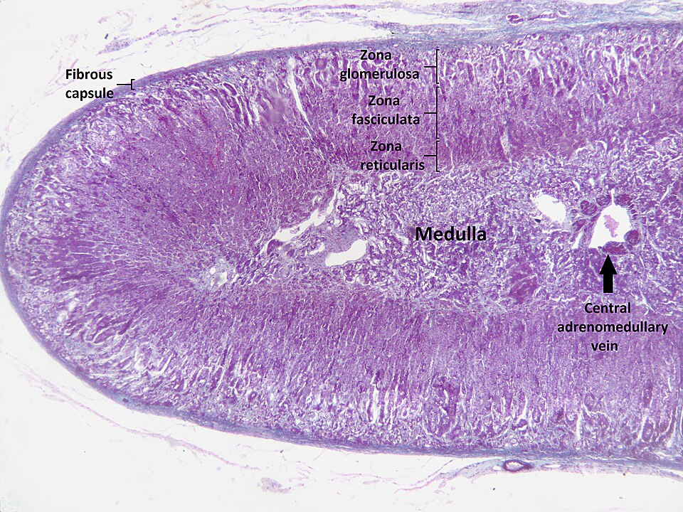
Light micrograph of a human adrenal gland with zones labeled: glomerulosa, fasciculata, reticularis, and the central medulla. This helps students relate the textbook diagram to actual tissue architecture. Extra labels (e.g., central vein) are included for orientation but are not required beyond recognising the main zones. Source.
Integration of Cortex and Medulla in Stress Response
The adrenal glands enable a dual stress response:
The medulla provides a rapid, nervous response to acute stress.
The cortex provides a slower, hormonal response to chronic stress.
Acute stress (short-term):
Sympathetic stimulation activates the adrenal medulla.
Adrenaline and noradrenaline prepare the body for immediate physical activity.
Chronic stress (long-term):
Hypothalamic release of corticotropin-releasing hormone (CRH) stimulates ACTH secretion.
ACTH maintains high cortisol levels, sustaining glucose availability for prolonged energy demands.
This integrated system ensures that the body can respond effectively to varying types and durations of stress.
Summary of Hormones and Their Functions
Adrenal Cortex:
Zona glomerulosa: Aldosterone — regulates sodium, potassium, and water balance.
Zona fasciculata: Cortisol — manages metabolism, glucose synthesis, and long-term stress.
Zona reticularis: Androgens — support secondary sexual characteristics and growth.
Adrenal Medulla:
Adrenaline: Increases heart rate, respiration, and blood glucose; prepares body for action.
Noradrenaline: Elevates blood pressure and maintains vascular tone.
Practice Questions
Question 1 (2 marks)
Name the two main regions of the adrenal gland and state one hormone produced by each.
Mark Scheme:
1 mark: Correctly names the two main regions as the adrenal cortex and adrenal medulla.
1 mark: Provides one correct hormone from each region:
Cortex: e.g. aldosterone, cortisol, or androgens.
Medulla: adrenaline or noradrenaline.
(1 mark for each correct region-hormone pair, maximum 2 marks)
Question 2 (5 marks)
Describe how the adrenal cortex and adrenal medulla help the body to respond to stress.
Mark Scheme:
1 mark: States that both regions of the adrenal gland are involved in the stress response but act through different mechanisms (hormonal vs nervous).
1 mark: Describes that the adrenal cortex releases steroid hormones (glucocorticoids such as cortisol) that regulate long-term responses to stress.
1 mark: Explains that cortisol increases blood glucose through gluconeogenesis and promotes protein and fat breakdown.
1 mark: States that the adrenal medulla releases adrenaline and noradrenaline under control of the sympathetic nervous system.
1 mark: Explains their short-term effects, such as increased heart rate, blood pressure, and blood glucose, preparing the body for “fight or flight.”
(Total 5 marks — award 1 mark for each distinct correct point)
FAQ
The adrenal cortex is composed of layers of polygonal epithelial cells arranged in cords or clusters, each layer having distinctive lipid content and staining patterns due to steroid synthesis.
Zona glomerulosa: Cells form rounded clusters; limited smooth endoplasmic reticulum for mineralocorticoid production.
Zona fasciculata: Large, lipid-rich cells in columns; abundant smooth endoplasmic reticulum for glucocorticoid synthesis.
Zona reticularis: Smaller cells with dense cytoplasm producing androgens.
The medulla, by contrast, contains chromaffin cells — modified neurones rich in granules of catecholamines and innervated by sympathetic fibres.
Blood enters via the adrenal arteries into a subcapsular plexus, then flows through sinusoids in the cortex before draining into the medulla.
This arrangement allows cortical hormones (like cortisol) to reach the medulla directly, influencing adrenaline synthesis by inducing phenylethanolamine N-methyltransferase (PNMT).
Finally, blood drains into the central adrenomedullary vein, enabling rapid hormone release into systemic circulation during stress.
Cortisol and adrenaline act synergistically to maintain short-term and long-term adaptation.
Adrenaline provides an immediate response, mobilising glucose and oxygen for muscle activity.
Cortisol sustains energy supply by promoting gluconeogenesis and inhibiting non-essential processes such as immune responses.
Cortisol also upregulates adrenaline receptor sensitivity, ensuring tissues remain responsive to sympathetic stimulation.
Aldosterone release is primarily regulated by the renin–angiotensin–aldosterone system (RAAS).
Low blood pressure or sodium triggers renin release from the kidney.
Renin converts angiotensinogen to angiotensin I, then to angiotensin II, which stimulates aldosterone secretion.
Aldosterone increases sodium reabsorption and water retention, restoring blood volume and pressure.
A smaller regulatory influence comes from increased plasma potassium, which directly stimulates aldosterone release.
Chromaffin cells are modified postganglionic sympathetic neurones that secrete adrenaline and noradrenaline rather than releasing neurotransmitters at a synapse.
They synthesise catecholamines from tyrosine through a series of enzymatic steps involving DOPA, dopamine, and noradrenaline.
In response to sympathetic stimulation, acetylcholine from preganglionic fibres triggers exocytosis of hormone-filled vesicles, releasing catecholamines into the bloodstream for rapid body-wide effects.
