Core idea: cells as the basic unit of life
Cells are the basic structural unit of all living organisms.
Cell theory predicts that any newly discovered organism should consist of one or more cells.
All cells need structures that allow genetic control, metabolism, exchange with surroundings, and internal organization.
Microscopy skills
Be able to make temporary mounts of cells and tissues.
Use stains to improve contrast and make structures easier to identify.
Focus using coarse adjustment first, then fine adjustment.
Measure specimens using an eyepiece graticule.
Calculate actual size and magnification.
Be able to produce a scale bar and take photographs.
Microscopy measurements are a form of quantitative observation.
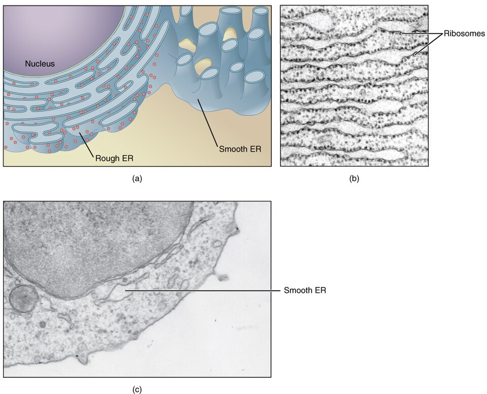
This image shows how microscopy can reveal cell ultrastructure that is not visible clearly with basic light microscopy. It is useful for linking structure to function, especially the difference between rough ER and smooth ER. Source
Developments in microscopy
Electron microscopy has much higher resolution and magnification than light microscopy, so it reveals ultrastructure.
Freeze fracture splits membranes along the hydrophobic interior, helping show membrane structure.
Cryogenic electron microscopy (cryo-EM) allows biological structures to be viewed in a more natural state.
Fluorescent stains can label specific cell components.
Immunofluorescence uses antibodies linked to fluorescent markers to locate specific proteins.
Structures common to all living cells
All typical cells contain DNA as genetic material.
All have cytoplasm, composed mainly of water, where many reactions occur.
All are enclosed by a plasma membrane made largely of lipids.
These shared structures support inheritance, metabolism, and controlled exchange.
Prokaryote cell structure
Required example: Gram-positive eubacteria such as Bacillus and Staphylococcus.
Main components: cell wall, plasma membrane, cytoplasm, naked DNA in a loop, and 70S ribosomes.
Prokaryotes do not have a nucleus.
Prokaryotes do not have membrane-bound organelles.
Prokaryotic structure varies, but detailed exceptions are not required.
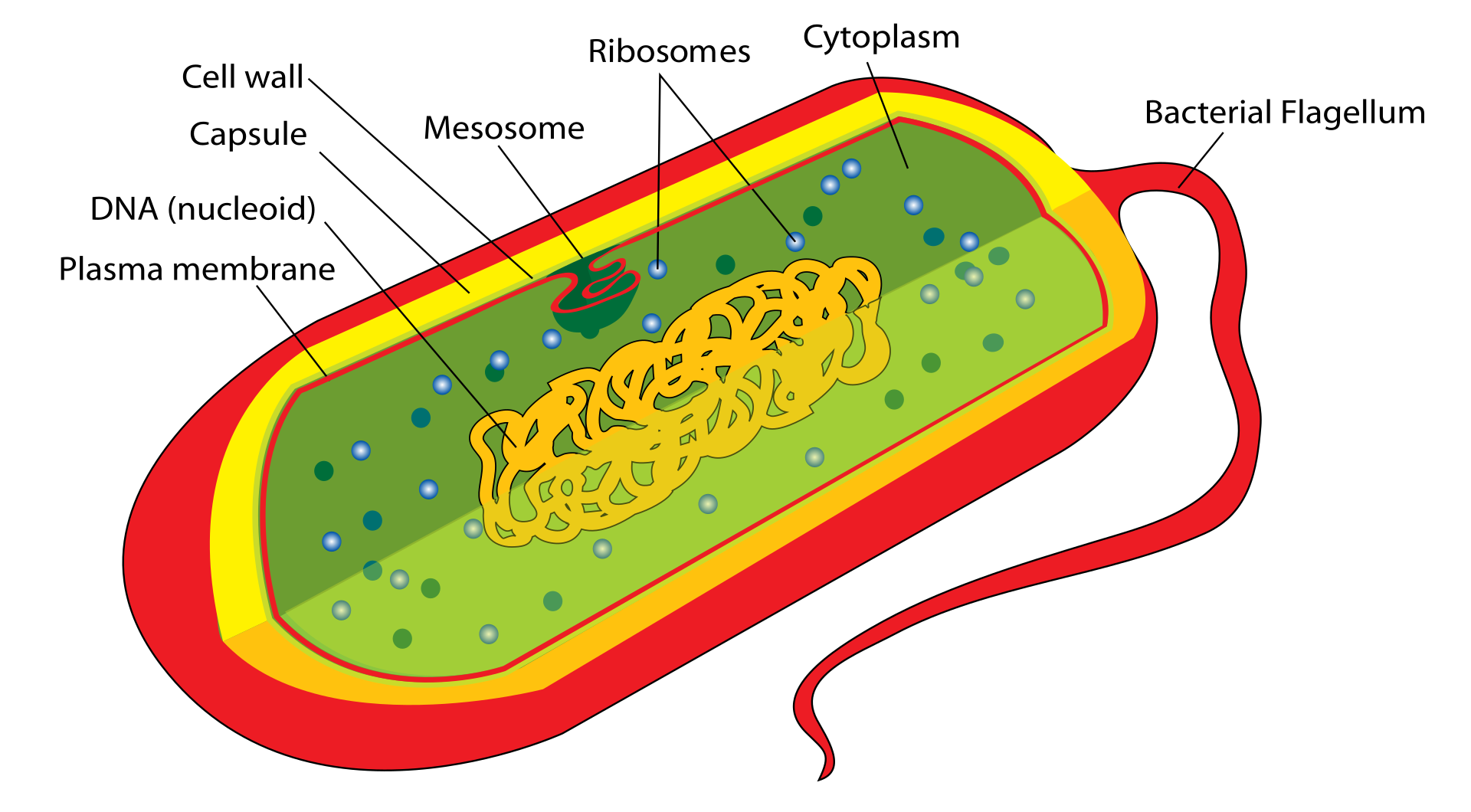
This image is useful for revising the simple organization of prokaryotes and contrasting them with eukaryotic cells, which are compartmentalized. Source
Eukaryote cell structure
Eukaryotes have a plasma membrane enclosing a compartmentalized cytoplasm.
They contain 80S ribosomes.
The nucleus contains chromosomes made of DNA bound to histones.
The nucleus is enclosed by a double membrane with pores.
Membrane-bound organelles include mitochondria, endoplasmic reticulum, Golgi apparatus, and vesicles/vacuoles including lysosomes.
Eukaryotic cells also contain a cytoskeleton of microtubules and microfilaments.
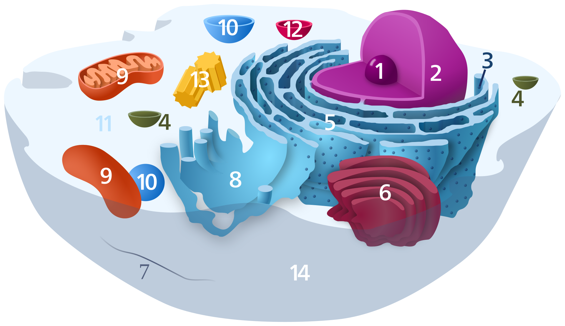
This image is useful for learning the layout of major eukaryotic organelles and linking labeled structure to function. Source
Life processes in unicellular organisms
A unicellular organism carries out all functions of life in one cell.
These include homeostasis, metabolism, nutrition, movement, excretion, growth, response to stimuli, and reproduction.
Exam idea: one cell can still perform the full range of life processes.
Differences between animal, fungal and plant cells
Cell walls differ:
Plants: cell wall present, made mainly of cellulose.
Fungi: cell wall present, but not cellulose-based like plants.
Animals: no cell wall.
Vacuoles differ in size and function.
Plants have chloroplasts and other plastids; animals and fungi do not.
Centrioles, cilia, and flagella are key distinguishing features in some eukaryotic cells.
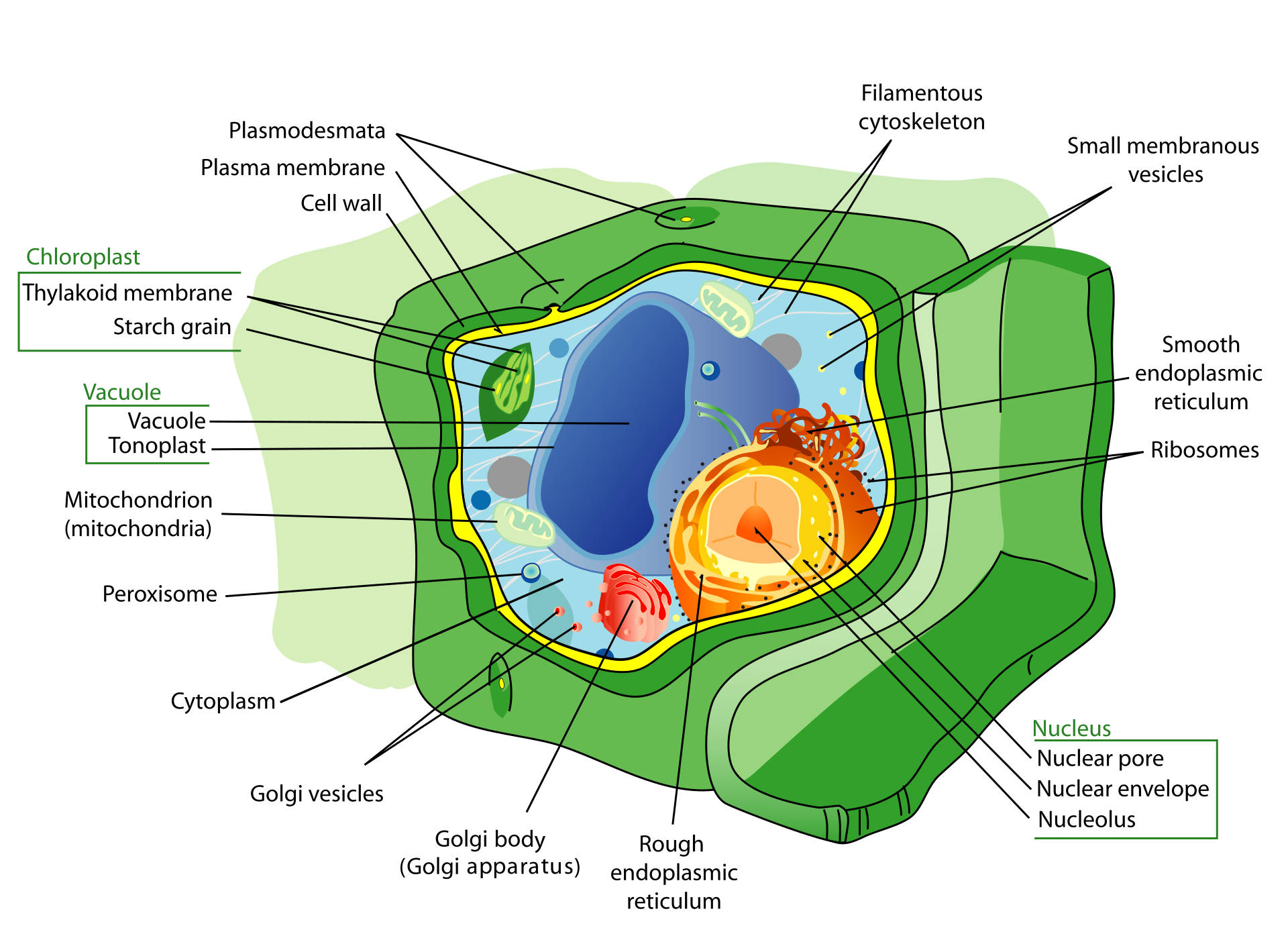
This image helps compare plant cells with animal cells and identify the key features that IB questions often test in diagrams and micrographs. Source
Atypical eukaryotic cells
Not all eukaryotic cells fit the “typical” model.
Use number of nuclei as the key comparison.
Aseptate fungal hyphae can have many nuclei in one continuous cell.
Skeletal muscle fibres are multinucleate.
Red blood cells in mammals lack a nucleus.
Phloem sieve tube elements also lack a nucleus at maturity.
Interpreting light and electron micrographs
Be able to identify a cell as prokaryotic, plant, or animal.
In electron micrographs, identify:
nucleoid region
prokaryotic cell wall
nucleus
mitochondrion
chloroplast
sap vacuole
Golgi apparatus
rough ER and smooth ER
chromosomes
ribosomes
cell wall
plasma membrane
microvilli
Use visible clues such as double membranes, internal membranes, size, and presence/absence of a nucleus.
This image is useful for practising how organelles appear in an electron micrograph, rather than as a textbook cartoon. Source
Drawing and annotation from electron micrographs
Be able to draw and annotate organelles seen in electron micrographs.
Required organelles: nucleus, mitochondria, chloroplasts, sap vacuole, Golgi apparatus, rough ER, smooth ER, chromosomes.
Required other structures: cell wall, plasma membrane, secretory vesicles, microvilli.
In annotations, include function as well as name.
Keep drawings clear, simple, and based only on what is visible.
HL only: origin of eukaryotic cells by endosymbiosis
Eukaryotes likely evolved from a common unicellular ancestor that already had a nucleus and reproduced sexually.
Mitochondria evolved by endosymbiosis.
In some eukaryotes, chloroplasts also evolved by endosymbiosis.
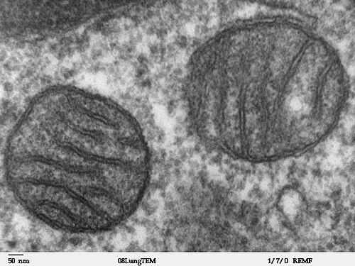
Evidence for endosymbiosis:
70S ribosomes inside mitochondria and chloroplasts
naked circular DNA
ability to replicate independently
The theory is strong because it explains many observations and makes accurate predictions.
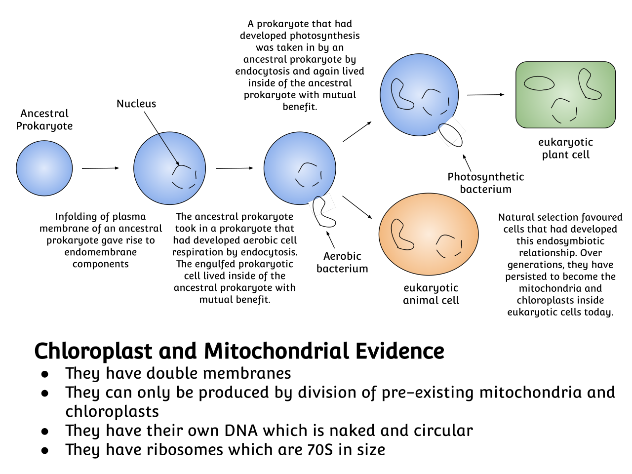
This image helps explain why these organelles share features with prokaryotes, including their own DNA, 70S ribosomes, and replication. Source
HL only: cell differentiation and multicellularity
Cell differentiation produces specialized tissues in multicellular organisms.
It is based on different patterns of gene expression.
Differentiation is often triggered by changes in the environment.
Multicellularity evolved repeatedly.
Many fungi, many eukaryotic algae, and all plants and animals are multicellular.
Advantages of multicellularity include larger body size and cell specialization.
Checklist: can you do this?
Distinguish between prokaryotic and eukaryotic cells using a diagram or micrograph.
Identify major organelles and cell structures in light and electron micrographs.
Calculate actual size and magnification, and add a correct scale bar.
Compare animal, plant, and fungal cell structure accurately.
Explain the evidence for endosymbiosis and link organelle features to the theory.
Fast exam traps to avoid
Do not say prokaryotes have a nucleus or membrane-bound organelles.
Do not confuse 70S ribosomes (prokaryotes, mitochondria, chloroplasts) with 80S ribosomes (eukaryotic cytoplasm).
Do not label every plant cell space as a vacuole unless it is clearly the sap vacuole.
Do not copy stylized textbook diagrams when asked to draw from a micrograph; only include what is visible.
Do not forget that some eukaryotic cells are atypical and may lack a nucleus or have many nuclei.