
AP Syllabus focus:
‘In a reflex arc, sensory neurons, interneurons, and motor neurons work together across the central and peripheral nervous systems to respond to stimuli.’
Reflexes show how the nervous system can produce rapid, automatic behaviour without conscious decision-making. Understanding reflex arcs and neuron types helps explain how information flows between the peripheral and central nervous systems.
Reflex arcs: the basic idea
A reflex is an involuntary, immediate response to a stimulus that promotes protection and efficient functioning.
Reflex arc: a neural pathway that carries information from a stimulus to a rapid response, typically with minimal involvement of higher brain centres.
Reflex arcs emphasise speed: they prioritise quick action over detailed analysis, which is why many are coordinated at the spinal cord level.
Core components of a reflex arc
Most reflex arcs include:
A receptor (detects the stimulus, e.g., pain, stretch)
A sensory neuron that brings information into the CNS
One or more interneurons within the CNS (often the spinal cord)
A motor neuron that sends commands out to muscles or glands
An effector (the muscle or gland producing the response)
Types of neurons in reflex pathways
Neurons are specialised cells that transmit information; reflex arcs mainly rely on three functional types.
Sensory neuron (afferent neuron): a neuron that carries incoming information from receptors in the body to the central nervous system.
Sensory neurons enter the CNS and provide the “input” signal that something has changed in the environment or body.
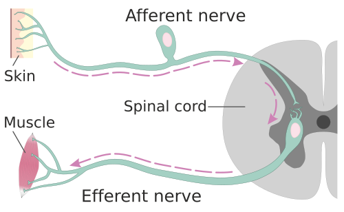
This schematic highlights the directional logic of reflex pathways: sensory (afferent) neurons carry information from receptors toward the spinal cord, while motor (efferent) neurons carry commands outward to effectors. It helps anchor the “input vs. output” distinction that underlies how reflex arcs coordinate rapid responses. Source
Interneurons (association neurons)
Interneurons are located entirely within the CNS and act as connectors and processors in reflexes. They can:
Relay signals from sensory neurons to motor neurons
Integrate information (e.g., amplify, inhibit, or coordinate responses)
Support more complex, polysynaptic reflexes involving multiple synapses
Interneuron: a CNS neuron that processes and transmits information between sensory and motor neurons, often enabling coordination and modulation of a response.
Interneurons are especially important when a reflex requires coordination across several muscles rather than a single, simple movement.
Motor neurons
Motor neurons carry commands from the CNS to effectors to produce movement or secretion.
Motor neuron (efferent neuron): a neuron that carries outgoing signals from the central nervous system to muscles or glands (effectors).
Motor neuron activation produces the observable reflex response, such as muscle contraction or withdrawal.
How reflex arcs cross the PNS and CNS
The syllabus emphasis is that reflex arcs span both systems:
The PNS detects stimuli and carries signals to/from the CNS
Sensory neurons travel from receptors toward the spinal cord/brain
Motor neurons travel from the spinal cord/brain to effectors
The CNS (often the spinal cord in simple reflexes) coordinates the response
Interneurons (when present) sit inside the CNS and link input to output
This division of labour allows rapid responding while still enabling the brain to receive information about what happened.
Key variations: mono- vs polysynaptic reflexes
Reflex arcs differ in the number of synapses involved:
Monosynaptic reflex: sensory neuron synapses directly onto a motor neuron
Typically very fast and stereotyped
Polysynaptic reflex: one or more interneurons between sensory and motor neurons
Often more flexible and can coordinate multiple effectors
Even when a reflex is coordinated in the spinal cord, sensory information can still ascend to the brain, supporting awareness and later voluntary adjustment without delaying the initial reflex response.
Practice Questions
State two functional types of neuron involved in a reflex arc and describe each role briefly. (2 marks)
1 mark: sensory (afferent) neuron carries information from receptor to CNS.
1 mark: motor (efferent) neuron carries command from CNS to effector (muscle/gland).
(Interneuron description may credit either point if clearly correct.)
Explain how a reflex arc demonstrates interaction between the peripheral and central nervous systems, referring to sensory neurons, interneurons, and motor neurons. (5 marks)
1 mark: identifies PNS role in carrying sensory input from receptors to CNS (sensory/afferent pathway).
1 mark: identifies CNS (often spinal cord) as a coordination site for reflexes.
1 mark: explains interneurons are within CNS and link/process between sensory and motor neurons (in polysynaptic reflexes).
1 mark: identifies PNS role in carrying output from CNS to effectors (motor/efferent pathway).
1 mark: links this organisation to rapid, automatic responding (minimal higher brain involvement at initiation).
FAQ
Yes. Descending signals from the brain can enhance or inhibit spinal reflex circuits, shifting response strength with attention, stress, expectation, or training.
Interneurons add flexibility. They allow coordination across multiple muscles and enable inhibition/excitation patterns that a single sensory-to-motor synapse cannot provide.
Specific tendon reflexes map to particular spinal segments and peripheral nerves. Reduced, absent, or exaggerated reflexes can indicate peripheral nerve injury, spinal cord lesions, or altered CNS inhibition.
It is a circuit pattern where activation of a muscle is paired with inhibition of its antagonist muscle, typically mediated by inhibitory interneurons, producing smoother and more effective movement.
Because the wiring of the reflex pathway is relatively fixed, producing a consistent response to a given stimulus; variability usually reflects modulation by interneurons and higher CNS input rather than conscious choice.
