
AP Syllabus focus:
‘The retina transduces visual information, the blind spot shows incomplete retinal input, and accommodation by the lens focuses images on the retina.’
Vision begins as light is focused by the eye and converted into neural signals by the retina. Those signals travel through organised pathways to the brain, where visual perception is constructed and refined.
From light to a focused image
Getting light onto the retina
Light enters through the cornea (major initial bending of light) and passes through the pupil (opening that regulates light entry).
The lens fine-tunes focus so a clear image lands on the retina.
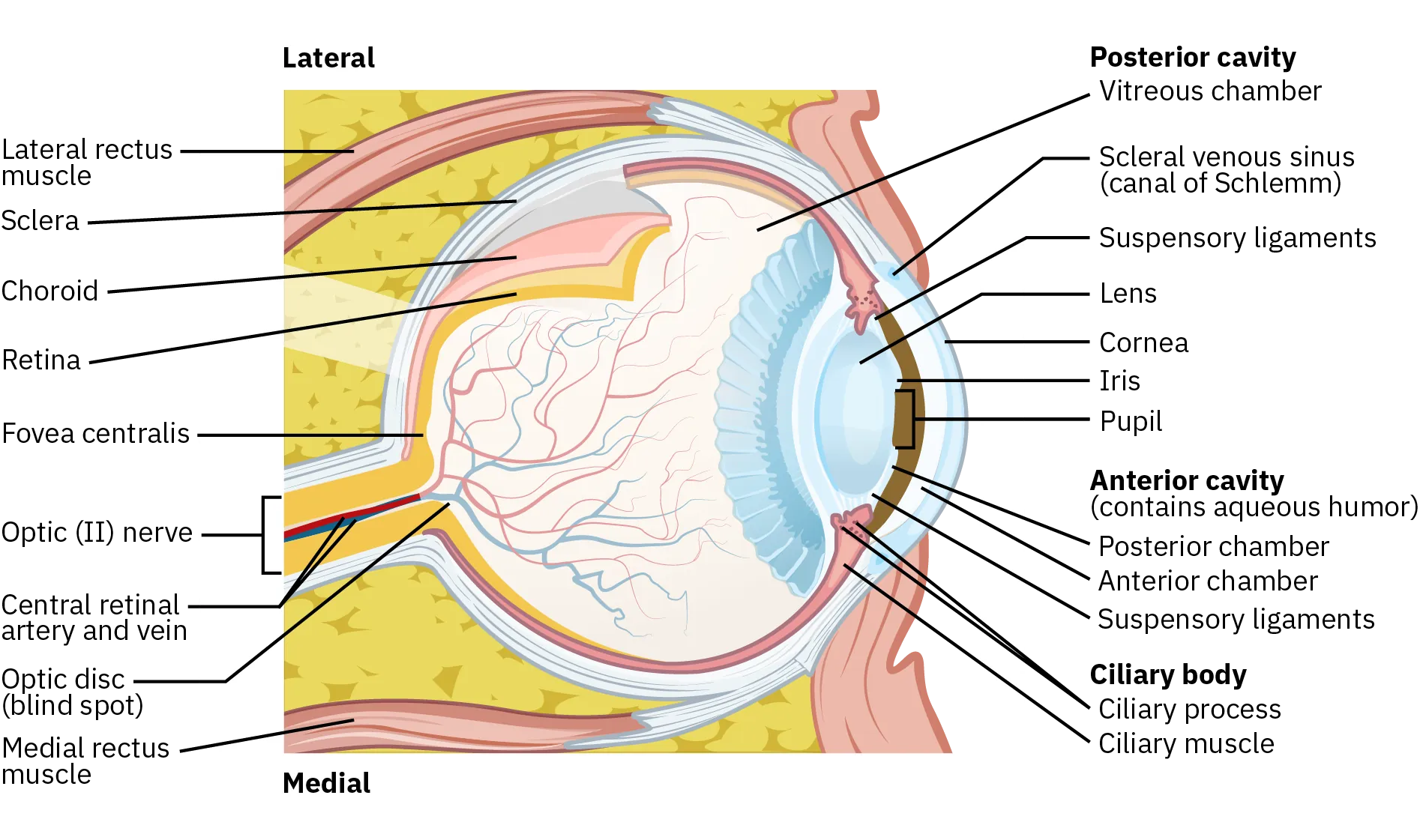
This cross-sectional eye anatomy diagram labels the cornea, pupil, lens, retina, and optic nerve, making the optical path of incoming light easy to visualize. It’s useful for connecting the optics (cornea/lens focusing) to the neural structures that begin visual processing (retina → optic nerve). Source
Accommodation (focusing up close vs far away)
Accommodation: the process by which the lens changes shape (via ciliary muscles) to focus near or far objects sharply on the retina.
Near objects: ciliary muscles contract → lens becomes more rounded → increases bending of light.
Far objects: ciliary muscles relax → lens flattens → decreases bending of light.
Accommodation supports visual clarity, especially when shifting gaze between distances (e.g., board to notebook).
The retina: converting light into neural signals
Retina as a transducer
The retina is a layered sheet of neural tissue lining the back of the eye. Its essential job is to transduce visual information—turning light energy into neural impulses the brain can process.
Transduction: the conversion of one form of energy into another; in vision, light energy is converted into neural signals.
Key pathway within the retina (conceptual flow):
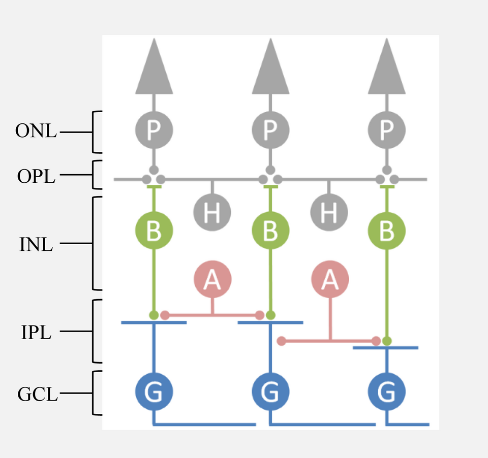
This diagram summarizes the retina’s layered circuit and the direction of information flow: photoreceptors capture light changes, bipolar cells relay signals, and ganglion cells send output via the optic nerve. It also highlights the modulatory roles of horizontal and amacrine cells, which shape contrast and timing before signals leave the eye.
Photoreceptors detect light changes and begin the signal.
Signals are relayed through intermediate retinal cells to ganglion cells.
Ganglion cell axons bundle to form the optic nerve, carrying information toward the brain.
The blind spot and “missing” retinal input
Where the optic nerve exits the retina is the optic disc, which contains no photoreceptors. This creates a blind spot: a small region of the visual field with incomplete retinal input.
The blind spot is usually unnoticed because:
The other eye often provides overlapping information.
The brain uses contextual filling-in, constructing a continuous scene from surrounding patterns.
From retina to brain: major relay points
Optic nerve to cortex (high-yield map)
After retinal processing, visual signals follow a well-organised route:
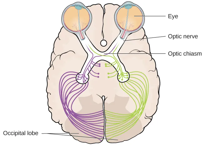
This illustration traces how visual information travels from the eyes through the optic nerves to the optic chiasm, where fibers partially cross so each hemisphere receives input from the contralateral visual field. It then shows the posterior route toward the occipital lobe, reinforcing the idea that perception depends on organized relays—not a single “wire” from eye to brain. Source
Optic nerve carries signals from each eye.
At the optic chiasm, some fibres cross, so each hemisphere receives information from the contralateral visual field.
Signals relay through the thalamus (especially the lateral geniculate region) and then project to the primary visual cortex in the occipital lobe, where basic features (like edges and orientation) are analysed.
Higher visual areas integrate these features into meaningful perception (objects, scenes), combining incoming input with prior knowledge and attention.
What “from retina to brain” implies for perception
The retina does early coding; perception is not a direct “copy” of the world.
Because the brain interprets and completes information (as seen with the blind spot), visual experience reflects both sensory input and neural processing.
Practice Questions
Define accommodation and state how it helps form a clear retinal image. (2 marks)
1 mark: Correct definition of accommodation (lens changes shape to focus).
1 mark: States that accommodation focuses near/far images sharply on the retina (clear retinal image).
A student closes their left eye and reports a small “gap” in vision when a dot is moved to a particular position while they fixate on a cross. Using psychological knowledge, explain the blind spot and outline the pathway of visual information from the retina to the visual cortex. (6 marks)
1 mark: Identifies the blind spot as the optic disc/point where optic nerve exits.
1 mark: States there are no photoreceptors at the blind spot (missing retinal input).
1 mark: Notes brain filling-in and/or binocular overlap reduces awareness.
1 mark: Retina transduces light into neural signals.
1 mark: Optic nerve carries signals; fibres partially cross at the optic chiasm (contralateral processing).
1 mark: Relay via thalamus to primary visual cortex in the occipital lobe.
FAQ
Neighbouring retinal cells inhibit one another’s activity, which heightens contrast at borders.
Strongly stimulated cells reduce neighbouring signals
Edges become more distinct than gradual changes in light
It helps organise and gate visual input before it reaches cortex.
It can prioritise signals based on alertness and attentional demands, influencing what gets processed most strongly downstream.
The optics of the cornea and lens invert the image on the retina.
The brain learns stable mappings between retinal patterns and the external world, so conscious perception is oriented correctly.
A receptive field is the region of the visual field that changes a neuron’s firing.
In early cortex, receptive fields support feature detection (e.g., line orientation), building blocks for later object perception.
Central retina has denser, more precisely connected circuitry supporting fine detail.
Peripheral regions pool information over larger areas, which boosts sensitivity but reduces acuity for small details.
