
IB Syllabus focus: 'Brain imaging techniques allow psychologists to investigate brain structure and activity non-invasively.'
Brain imaging has transformed biological psychology by letting researchers study the living brain safely. These methods reveal either brain structure or brain activity and provide evidence for explanations of behavior and cognition.
Why non-invasive brain research matters
Before modern imaging, evidence about the brain often came mainly from postmortem examination or accidental injury. Non-invasive methods changed this by allowing psychologists to collect data from healthy participants while the brain is intact and functioning. This makes it possible to compare groups, track change over time, and link patterns in the brain to performance on cognitive or behavioral tasks.
Non-invasive research: Research methods that examine the brain or body without surgery or physically entering tissue.
Non-invasive methods are especially valuable because they can usually be repeated, creating more opportunities for reliable measurement and replication. However, each technique measures a different aspect of the brain, so the choice of method strongly affects what researchers can conclude.
Structural and functional imaging
Psychologists usually distinguish between methods that show brain structure and methods that show brain activity.
Structural imaging: Techniques that produce images of brain anatomy. Functional imaging: Techniques that measure brain activity over time.
Structural methods are useful when researchers want to examine the size, shape, or condition of brain areas.
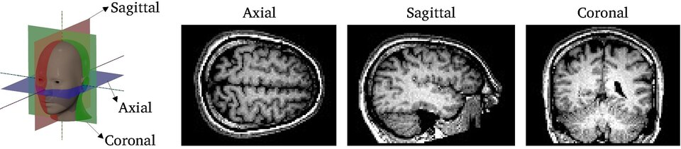
Diagram of the three main anatomical imaging planes (axial, sagittal, and coronal) used to view brain structure in MRI. Understanding these planes helps you interpret what “location” and “structure” mean when psychologists discuss MRI-based findings. Source
Functional methods are used when the goal is to investigate what the brain is doing during a task or at rest.
Common techniques
MRI (magnetic resonance imaging) creates detailed images of brain anatomy using strong magnetic fields and radio waves.
It produces high-quality images of soft tissue.
It is especially useful for identifying structural differences or abnormalities.
It has strong spatial resolution, meaning it can locate structures precisely.
fMRI (functional magnetic resonance imaging) measures changes in blood oxygenation, often called the BOLD signal.
Increased blood flow is treated as an indirect sign that neurons in an area were more active.
fMRI can show which brain regions are more active in different conditions.
It has good spatial resolution but weaker temporal resolution than EEG because blood-flow changes lag behind neural firing.
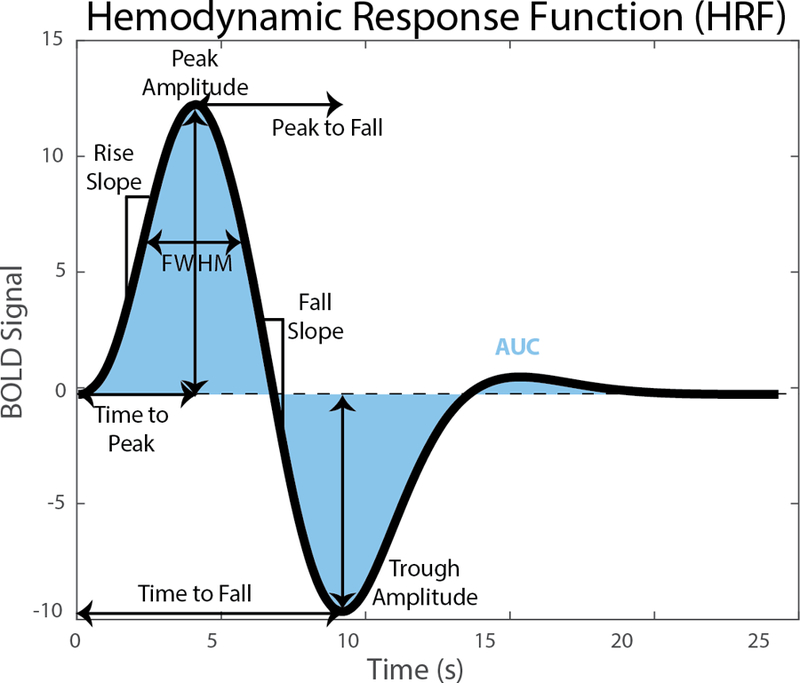
Example hemodynamic response function (HRF) showing how the fMRI BOLD signal changes over time after neural activity. The delayed peak (on the order of a few seconds) visualizes why fMRI timing is less precise than EEG, even though spatial localization is typically stronger. Source
EEG (electroencephalography) records electrical activity from the scalp using electrodes.
It is useful for showing the timing of brain processes in milliseconds.
This makes it highly valuable when researchers care about the sequence of processing.
Its main weakness is poor spatial resolution, so it is harder to identify the exact source of activity.
How psychologists evaluate imaging data
A major issue in brain imaging is the trade-off between where activity happens and when it happens. MRI and fMRI are stronger for identifying location. EEG is stronger for identifying timing. Because no single method is perfect, psychologists often choose the technique that best matches the research question.
Brain imaging data must also be interpreted carefully. Some methods, such as EEG, measure neural activity more directly. Others, such as fMRI, measure a related biological change rather than neural firing itself. This means that scans do not provide a simple picture of “the mind”; they provide evidence that must be analyzed statistically and interpreted in context.
Another important point is that imaging findings are often correlational. If a brain area is active during a task, this suggests an association between that area and the task, but it does not by itself prove that the activity caused the behavior. Strong claims are more convincing when imaging results are supported by other methods or replicated across studies.
Strengths of brain imaging and non-invasive methods
Non-invasive imaging has several major strengths for psychology:
It allows study of the living brain in real time or near real time.
It reduces the risks associated with surgical methods.
It can be used with healthy participants as well as many clinical groups.
It often produces objective, visual data that can be compared across participants.
It supports repeated testing, which is useful for longitudinal research or checking consistency.
These methods have helped make biological explanations in psychology more scientific because they allow direct observation of brain structure and measurable patterns of activity rather than relying only on speculation.
Limitations and cautions
Despite their value, brain imaging methods do not provide perfect evidence. Brain activity can be affected by movement, fatigue, anxiety, and the artificial setting of the scanner or laboratory. For example, MRI and fMRI environments can be noisy and restrictive, which may influence participant behavior.
Different methods also vary in cost and accessibility. MRI and fMRI are expensive and require specialized equipment and expertise, which can limit sample size. Small samples can reduce confidence in how widely findings apply.
There are also interpretation problems. Brightly colored brain images can appear very precise, but the final image usually reflects substantial processing and averaging. In addition, an active region may be only one part of a larger network. Overinterpreting a single scan can lead to simplistic claims.
Finally, psychologists must consider participant well-being and data sensitivity. Even non-invasive scans may cause discomfort, and brain data can reveal unexpected information. Researchers therefore need careful consent procedures, confidentiality, and clear plans for handling unusual findings.
Practice Questions
(2 marks): State one difference between MRI and fMRI.
1 mark for stating that MRI measures brain structure or anatomy.
1 mark for stating that fMRI measures brain activity indirectly through blood oxygenation or the BOLD signal.
(6 marks): Explain one strength and one limitation of using EEG in non-invasive psychological research.
1 mark for identifying EEG as a non-invasive brain imaging or recording technique.
2 marks for explaining one strength, such as excellent temporal resolution or the ability to detect timing of brain activity in milliseconds.
2 marks for explaining one limitation, such as poor spatial resolution or difficulty locating the exact source of activity.
1 mark for linking the strength and limitation to psychological research, such as studying the sequence of mental processing or limits on interpreting where activity occurs.
FAQ
MRI noise is produced by rapidly switching gradient coils, which vibrate during scanning.
This can matter in studies of hearing, attention, or stress because the sound may influence participant responses. Researchers often use ear protection, quieter scanning sequences, or control tasks designed to account for scanner noise.
EEG electrodes need good electrical contact with the scalp. Gel or saline lowers resistance at the skin surface and improves signal quality.
If contact is poor, the recording can become noisy or unstable. Better contact helps researchers detect real brain signals rather than interference from hair, skin, or small movements.
Preprocessing is the set of steps used to clean and prepare raw brain imaging data before analysis.
Depending on the method, this may include removing noise, correcting for motion, aligning scans, or averaging repeated signals. Without preprocessing, the results may reflect artifacts or technical errors instead of genuine brain structure or activity.
Raw fMRI activity reflects many things at once, including perception, movement, attention, and task difficulty.
Researchers therefore compare one condition with another to isolate the process they want to study. A good control condition helps narrow the interpretation of the BOLD signal and makes the findings more meaningful.
Some non-invasive methods detect signals best from areas near the surface of the brain. For example, EEG records from the scalp, so signals from deep structures can be weaker and harder to localize.
Even with fMRI, some deep or lower brain regions can be harder to measure clearly because of signal loss or distortion. This means findings may be stronger for some brain areas than others.
