
IB Syllabus focus: 'Brain imaging has contributed to understanding neuroplasticity and interactions between the brain and environment.'
Neuroplasticity shows that the brain is dynamic rather than fixed. Brain imaging research demonstrates that learning, stress, injury, and everyday experience can reshape brain structure and function across the lifespan.
Neuroplasticity
Neuroplasticity refers to the brain’s capacity to adapt rather than remain permanently unchanged.
Neuroplasticity: The brain’s ability to change its structure, organization, or functioning in response to experience, learning, development, or injury.
Plasticity can involve strengthened or weakened synapses, growth of new neural connections, pruning of unused connections, and reorganization of brain activity. This means behavior is not produced by a completely fixed biological system. Instead, the brain is continually modified by what a person repeatedly does, thinks, and experiences.
Plasticity does not mean unlimited change. Some systems are more flexible than others, and change is usually shaped by repetition, attention, age, intensity of experience, and how long a skill or condition lasts.
Main forms of plastic change
Structural changes involve measurable differences in brain matter, connections, or volume.
Functional reorganization happens when patterns of activation shift, such as after practice or after injury.
Use-dependent change means neural circuits become stronger when they are repeatedly activated and weaker when they are neglected.
Brain-Environment Interaction
The idea of brain-environment interaction emphasizes a two-way relationship: environments influence the brain, and the brain influences how the environment is perceived and managed.
Brain-environment interaction: The reciprocal relationship in which experiences and surroundings affect brain development and functioning, while the brain shapes responses to those experiences and surroundings.
This interaction helps explain why different people can show different neural patterns after different life experiences. It also explains why the same environment may not affect everyone in exactly the same way.
Common environmental influences
Learning and practice can strengthen neural pathways involved in memory, movement, language, or spatial skills.
Occupational demands may lead to long-term adaptation in brain areas used heavily in daily work.
Stress or adversity can alter brain systems linked to memory, emotion, and regulation.
Rehabilitation and stimulation after injury can encourage the brain to reorganize and support recovery.
Brain Imaging Evidence
Brain imaging has been especially important because psychologists can study living brains non-invasively and, in some cases, track changes over time. Structural MRI is useful for detecting changes in brain volume or gray matter, while functional scans can show shifting patterns of activity.
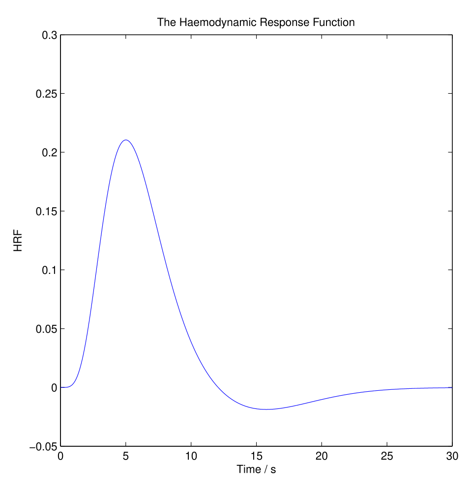
Canonical hemodynamic response function (HRF) used in fMRI analyses to model the BOLD signal over time. The curve highlights that fMRI ‘activation’ reflects a delayed vascular response (rise, peak, undershoot) rather than a direct, instantaneous recording of neuronal firing. Source
Together, these methods have made neuroplasticity visible rather than just theoretical.
Maguire et al. (2000)
A well-known study by Maguire et al. examined London taxi drivers, whose work requires extensive spatial navigation. MRI scans showed that the drivers had a larger posterior hippocampus than control participants.
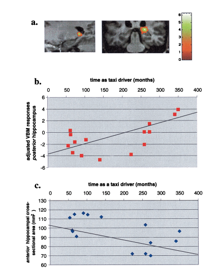
PNAS figure from Maguire et al. (2000) showing hippocampal gray-matter differences in London taxi drivers and how posterior hippocampal measures correlate with months/years of taxi-driving experience. The plots illustrate the core IB point that sustained environmental demands (navigation practice) can be associated with measurable structural brain changes. Source
The hippocampus is strongly associated with spatial memory, so the findings suggested that long-term navigation demands were linked to structural brain change.
The study also found a positive correlation between years of experience as a taxi driver and posterior hippocampal volume. This supports the idea that repeated environmental demands may shape the brain over time.
However, because the study was largely correlational, it cannot prove with certainty that taxi driving caused the change. It is possible that people with stronger spatial abilities were more likely to become taxi drivers in the first place.
Draganski et al. (2004)
Draganski et al. provided stronger evidence through a longitudinal design. Participants who learned to juggle were scanned before training, after learning, and again after a period without practice. MRI results showed increased gray matter in visual-motion areas after juggling was learned.
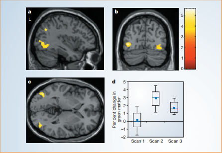
Figure 1 from Draganski et al. (2004) summarizing training-related structural plasticity during juggling practice. The statistical maps localize gray-matter increases in visual-motion regions (hMT/V5), and the accompanying plot shows the characteristic ‘increase then partial decrease’ pattern across Scan 1, Scan 2, and Scan 3 when practice stops. Source
When practice stopped, some of this increase decreased.
This study is important because it showed that:
structural brain change can occur in healthy adults
change can happen over a relatively short period
plasticity is linked to environmental demands and can be partly reversible
The study supports the idea that the brain responds directly to new skills and changing experience.
What Brain Imaging Has Shown
Taken together, imaging research suggests several important points.
Plasticity continues beyond childhood. Adult brains can still change in response to learning and experience.
The environment leaves measurable traces on the brain. Demanding, repeated, or meaningful experience can be linked to observable neural differences.
Plasticity is dynamic. Some changes are maintained with continued use, while others weaken when the relevant activity stops.
Brain change can be adaptive. Reorganization may support learning, expertise, or recovery after damage.
Evaluating the Evidence
Brain imaging has improved psychological understanding of neuroplasticity, but findings must be interpreted carefully.
Strength: Imaging allows repeated, non-invasive study of living brains, which is essential for tracking change over time.
Strength: Longitudinal designs provide stronger evidence for environmental influence than one-time comparisons.
Limitation: Imaging often shows association rather than direct cause. A visible brain difference does not always show exactly how the change happened.
Limitation: Structural change does not automatically mean better performance. Behavioral evidence is needed alongside imaging data.
Limitation: Many studies use small or specialized samples, so findings may not generalize equally to all people or all environments.
Practice Questions
State what is meant by neuroplasticity. [2 marks]
1 mark for identifying that the brain can change or adapt.
1 mark for stating that the change is in structure, organization, or function due to experience, learning, development, or injury.
Explain one brain imaging study that demonstrates neuroplasticity and brain-environment interaction. [6 marks]
1-2 marks for accurate description of a relevant study, such as Maguire et al. or Draganski et al.
1-2 marks for accurate findings from the brain imaging data.
1-2 marks for clearly linking the findings to neuroplasticity and showing how environmental experience affected the brain.
FAQ
Yes. Childhood is generally a period of greater plasticity because the brain is still developing rapidly.
However, adult brains are still capable of meaningful change. Research on skill learning, rehabilitation, and habit formation shows that adulthood involves ongoing plasticity, even if it is often slower or more dependent on repeated practice.
Maladaptive plasticity is brain change that occurs, but in an unhelpful way.
Examples can include:
chronic pain pathways becoming more strongly established
traumatic responses becoming deeply conditioned
inefficient habits becoming automatic through repetition
So, plasticity is not always positive. It simply means the brain changes.
Sleep supports the stabilization of new learning. After practice or study, the brain continues processing information during sleep.
This can help:
strengthen recently activated neural pathways
improve memory consolidation
support more efficient future performance
Without enough sleep, plastic changes linked to learning may be weaker.
Often, yes. Although earlier intervention is usually helpful, later rehabilitation can still produce change.
Improvement depends on factors such as:
the area and extent of damage
the type of therapy
intensity and consistency of practice
motivation and support in the environment
This is one reason rehabilitation programs often emphasize repeated, meaningful activity.
With repetition, the brain can process a skill more efficiently. Fewer attentional resources may be needed because the relevant neural circuits become more established.
This does not mean the brain is “working less.” It means processing has become more organized and efficient through plastic change.
Examples include reading familiar words, typing, or performing a well-learned motor routine.
